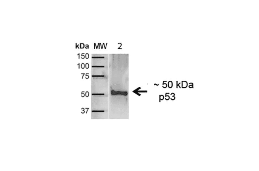
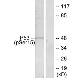
Western blot analysis of human A431 showing detection of ~43.7kDa p53 protein using Anti-p53 Antibody (A304903) at 1:1,000 for 1 hour at room temperature. Lane 1: MW Ladder. Lane 2: human A431 (20 µg). Load: 20 µg. Block: 5% milk + TBST for 1 hour at room temperature. The secondary antibody used was Goat Anti-Rabbit: HRP at 1:2000 for 1 hour at room temperature. Color Development: TMB solution for 12 min at room temperature. Predicted/Observed Size: ~43.7kDa.
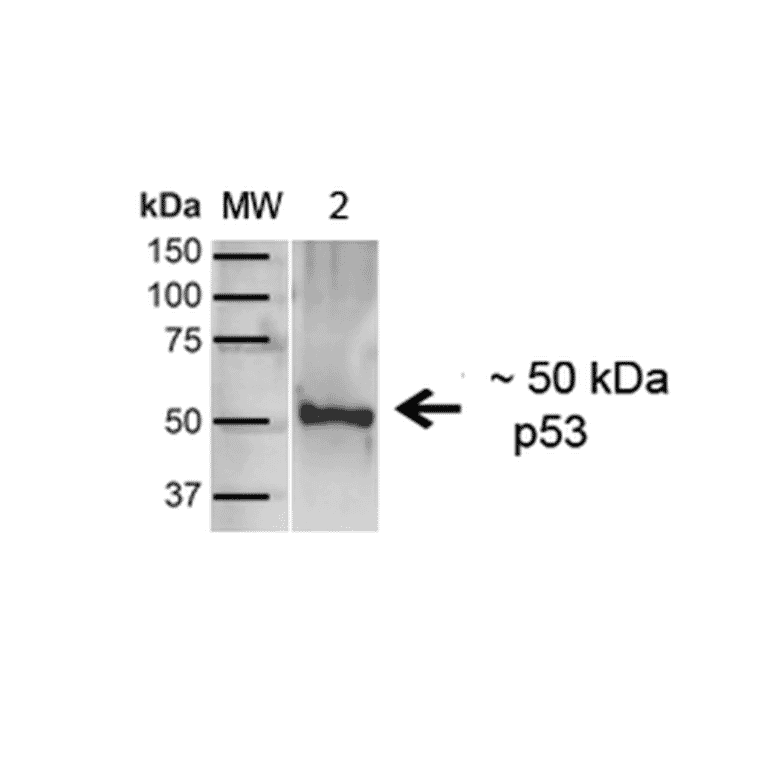
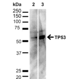
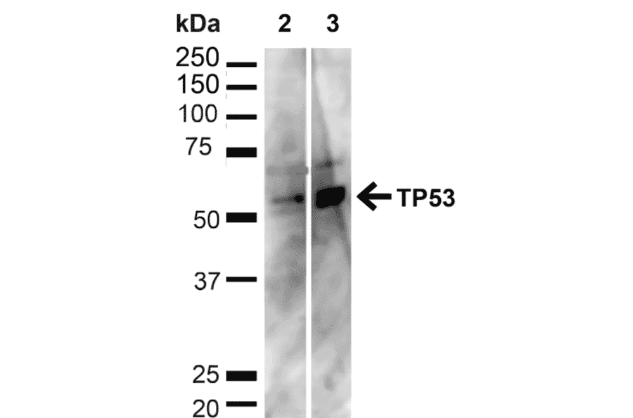
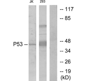
Western blot analysis of human HeLa and HEK293T cell lysates showing detection of ~43.7kDa p53 protein using Anti-p53 Antibody (A304903) at 1:1,000 for 1 hour at room temperature. Lane 1: MW Ladder. Lane 2: human HeLa (20 µg). Lane 3: human 293T (20 µg). Load: 20 µg. Block: 5% milk + TBST for 1 hour at room temperature. The secondary antibody used was Goat Anti-Rabbit: HRP at 1:2000 for 1 hour at room temperature. Color Development: TMB solution for 12 min at room temperature. Predicted/Observed Size: ~43.7kDa.
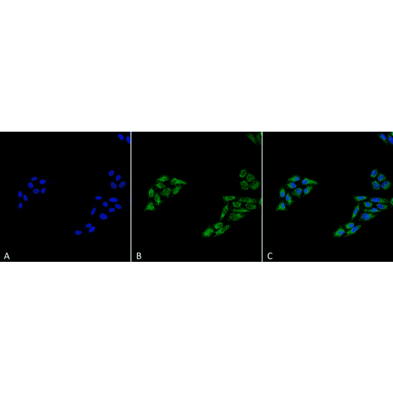
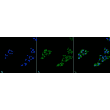
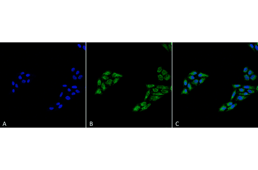
Immunocytochemistry/Immunofluorescence analysis of human cervical cancer cell line (HeLa), fixed in 4% formaldehyde for 15 min at room temperature, using Anti-p53 Antibody (A304903), at 1:100 for 60 minutes at room temperature. The secondary antibody used was Goat Anti-Rabbit ATTO 488 at 1:100 for 60 minutes at room temperature. Counterstain: DAPI (blue) nuclear stain at 1:5,000 for 5 minutes room temperature. Localization: Cytoplasm, PML body, Endoplasmic Reticulum. Magnification: 40X.(A) DAPI (blue) nuclear stain (B) Phalloidin Texas Red F-Actin stain (C) p53 Antibody (D) Composite.
Publishing research using Anti-p53 Antibody (A304903)? Please let us know so that we can list the citation on this page.
Alternative products to Anti-p53 Antibody (A304903)


![Immunocytochemistry - Anti-p53 Antibody [BP53-12] (A86030) - Antibodies.com](https://cdn.antibodies.com/image/catalog/86/A86031_387.jpg?profile=product_alternative)

![Immunohistochemistry - Anti-p53 (phospho Ser392) Antibody [FP3.2 [FPS392]] (A86708) - Antibodies.com](https://cdn.antibodies.com/image/catalog/86/A86708_827.jpg?profile=product_alternative)
![Immunohistochemistry - Anti-p53 Antibody [rBP53-12] (A250176) - Antibodies.com](https://cdn.antibodies.com/image/catalog/250/A250176_1.jpg?profile=product_alternative)