This antibody reacts with vimentin, a 57 kDa intermediate filament expressed in variety of mesenchymal and mesodermal cell types. Cross-reactivity was found with smooth muscle desmin.
Applications
IHC-P, WB, ICC, Flow Cytometry
Dilutions
IHC: Staining technique: (a) fix cells for 10 min in methanol at -20°C and for 6 min in acetone at -20°C; (b) fix cells directly in methanol for 10 min at -20°C or in acetone for 10 min at -20°C. Positive Control: 3T3 murine Swiss albino fibroblast cell line, RBL rat basophilic leukemia cell line, Flow Cytometry: 1-5 µg/ml; Intracellular staining.
Reactivity
Mammalian
Immunogen
Pellet of porcine brain cold-stable proteins after depolymerization of microtubules.
Host
Mouse
Clonality
Monoclonal
Clone ID
VI-01
Isotype
IgM
Conjugate
Unconjugated
Purification
Purified by differential precipitation and solid-phase chromatography.
Concentration
1 mg/ml
Predicted MW
57 kDa
Product Form
Liquid
Formulation
Supplied in Tris Buffered Saline, pH 8, with 15 mM Sodium Azide.
Separation of HeLa cells stained with Anti-Vimentin Antibody [VI-01] (A86296), (concentration in sample 5 µg/ml, GAM APC, red-filled) from HeLa cells unstained by primary antibody (GAM APC, black-dashed) in flow cytometry analysis (intracellular staining of methanol permeabilisated cells).
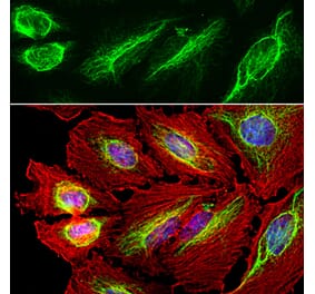
Immunohistochemistry staining (paraffin sections) of vimentin in human liver using Anti-Vimentin Antibody [VI-01] (A86296), (diluted 1:400), detected with GAM IgM-Alexa Fluor®488 (diluted 1:200; green), cell nuclei stained with PI (1µg/ml; orange).
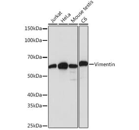
Anti-Vimentin Antibody [VI-01] (A86296) works in WB application under both reducing and non-reducing conditions. Western blotting analysis was performed on whole cell extracts (RIPA lysis buffer) of JAR, JEG3, HTr-8/SVneo, and HeLa cell lines mixed and heated (100°C, 5 min) with reducing (2-mercaptoethanol) or non-reducing SDS-loading buffer. Samples were resolved using 10% Tris-glycine SDS gel electrophoresis. Nitrocellulose membrane blot was probed simultaneously with mouse IgM monoclonal antibody VI-01 (1 µg/ml), and mouse IgG1 anti-tubulin monoclonal antibody TU-01 (1 µg/ml) used as the loading control. Subclass-specific secondary antibodies IRDye 800CW Goat-anti-Mouse IgG (green) and IRDye 680RD Goat-anti-Mouse IgM (red) were used for multiplex fluorescent Western blot detection. Vimentin was detected at ~55 kDa.
Immunoprecipitation of vimentin from HeLa cell lysate by Anti-Vimentin Antibody [VI-10] (A86652) and its detection by Anti-Vimentin Antibody [VI-01] (A86296). IgM heavy chain (76-92 kDa) and IgM light chain (25-30 kDa) indicated. MW of vimentin is 57 kDa. Lr = Lysate (reducing conditions). Lnr = Lysate (non-reducing conditions). IPr = Immunoprecipitate (reducing conditions). IPnr = Immunoprecipitate (non-reducing conditions).


![Flow Cytometry - Anti-Vimentin Antibody [VI-01] (A86296)](https://cdn.antibodies.com/image/catalog/86/A86296_1.jpg?profile=product_top)
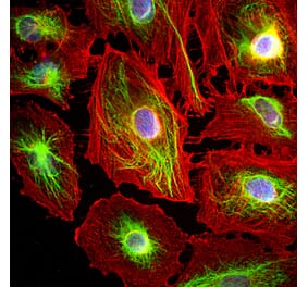
![ICC - Anti-Vimentin Antibody [VI-01] (A86296)](https://cdn.antibodies.com/image/catalog/86/A86296_2.jpg?profile=product_top)
![IHC - Anti-Vimentin Antibody [VI-01] (A86296)](https://cdn.antibodies.com/image/catalog/86/A86296_3.png?profile=product_top)
![WB - Anti-Vimentin Antibody [VI-01] (A86296)](https://cdn.antibodies.com/image/catalog/86/A86296_4.jpg?profile=product_top)
![IP - Anti-Vimentin Antibody [VI-01] (A86296)](https://cdn.antibodies.com/image/catalog/86/A86296_5.jpg?profile=product_top)
![Flow Cytometry - Anti-Vimentin Antibody [VI-01] (A86296)](https://cdn.antibodies.com/image/catalog/86/A86296_1.jpg?profile=product_top_thumb)
![ICC - Anti-Vimentin Antibody [VI-01] (A86296)](https://cdn.antibodies.com/image/catalog/86/A86296_2.jpg?profile=product_top_thumb)
![IHC - Anti-Vimentin Antibody [VI-01] (A86296)](https://cdn.antibodies.com/image/catalog/86/A86296_3.png?profile=product_top_thumb)
![WB - Anti-Vimentin Antibody [VI-01] (A86296)](https://cdn.antibodies.com/image/catalog/86/A86296_4.jpg?profile=product_top_thumb)
![IP - Anti-Vimentin Antibody [VI-01] (A86296)](https://cdn.antibodies.com/image/catalog/86/A86296_5.jpg?profile=product_top_thumb)
![Flow Cytometry - Anti-Vimentin Antibody [VI-01] (A86296)](https://cdn.antibodies.com/image/catalog/86/A86296_1.jpg?profile=product_image)
![ICC - Anti-Vimentin Antibody [VI-01] (A86296)](https://cdn.antibodies.com/image/catalog/86/A86296_2.jpg?profile=product_image)
![IHC - Anti-Vimentin Antibody [VI-01] (A86296)](https://cdn.antibodies.com/image/catalog/86/A86296_3.png?profile=product_image)
![WB - Anti-Vimentin Antibody [VI-01] (A86296)](https://cdn.antibodies.com/image/catalog/86/A86296_4.jpg?profile=product_image)
![IP - Anti-Vimentin Antibody [VI-01] (A86296)](https://cdn.antibodies.com/image/catalog/86/A86296_5.jpg?profile=product_image)
![ICC - Anti-Vimentin Antibody [VI-10] (A86652)](https://cdn.antibodies.com/image/catalog/86/A86652_1.jpg?profile=product_alternative)
![Immunohistochemistry - Anti-Vimentin Antibody [RV202] (A115651) - Antibodies.com](https://cdn.antibodies.com/image/catalog/115/A115651_1.jpg?profile=product_alternative)
![Immunohistochemistry - Anti-Vimentin Antibody [VIM/1937R] - BSA and Azide free (A250314) - Antibodies.com](https://cdn.antibodies.com/image/catalog/253/A253494_1.jpg?profile=product_alternative)
![Immunohistochemistry - Anti-Vimentin Antibody [VIM/1937R] (A253488) - Antibodies.com](https://cdn.antibodies.com/image/catalog/250/A250314_1.jpg?profile=product_alternative)

![Immunohistochemistry - Anti-Vimentin Antibody [V9] (A252494) - Antibodies.com](https://cdn.antibodies.com/image/catalog/250/A250310_1.jpg?profile=product_alternative)
![Immunohistochemistry - Anti-Vimentin Antibody [V9] - BSA and Azide free (A250310) - Antibodies.com](https://cdn.antibodies.com/image/catalog/253/A253490_1.jpg?profile=product_alternative)
![Western Blot - Anti-Vimentin Antibody [VM452] (A253577) - Antibodies.com](https://cdn.antibodies.com/image/catalog/250/A250307_1.jpg?profile=product_alternative)
![Western Blot - Anti-Vimentin Antibody [VM452] - BSA and Azide free (A250307) - Antibodies.com](https://cdn.antibodies.com/image/catalog/253/A253487_1.jpg?profile=product_alternative)
![Immunohistochemistry - Anti-Vimentin Antibody [VIM/6576R] (A250313) - Antibodies.com](https://cdn.antibodies.com/image/catalog/250/A250313_1.jpg?profile=product_alternative)
![Immunohistochemistry - Anti-Vimentin Antibody [VIM/6576R] - BSA and Azide free (A253493) - Antibodies.com](https://cdn.antibodies.com/image/catalog/253/A253493_1.jpg?profile=product_alternative)