This antibody is specific for Arrestin 1 and does not cross-react with cone Arrestin, beta Arrestin 1, or beta Arrestin 2.
Anwendungen
WB, ICC/IF, IHC
Verdünnungen
WB: 1:5,000, ICC/IF: 1:1,000
Reaktivität
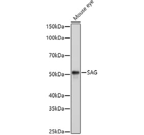
Human, Rat, Mouse, Bovine, Porcine, Horse
Immunogen
Recombinant bovine Arrestin 1, with the first 20 amino acids of the C-terminus truncated.
Wirt
Mouse
Klonalität
Monoclonal
Klon
S128
Isotyp
IgG1
Konjugat
Unconjugated
Reinigung
Immunogen affinity purification.
Konzentration
1 mg/ml
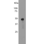
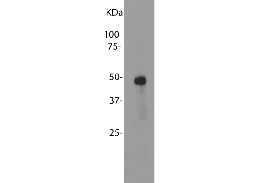
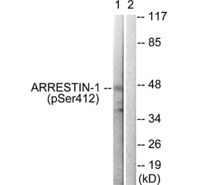
Molekulargewicht
48 kDa
Produktform
Liquid
Formulierung
Supplied in Phosphate Buffered Saline with 50% Glycerol and 5mM Sodium Azide.
Lagerung
Shipped at 4°C. Upon delivery aliquot and store at -20°C. Avoid freeze / thaw cycles.
Synonyme
48 kDa protein, Arrestin, ARRS_HUMAN, Retinal S antigen, Retinal S antigen (48 KDa protein), Retinal S-antigen, Rod photoreceptor arrestin, RP47, S antigen, S antigen retina and pineal gland (arrestin), S arrestin, S-AG, S-arrestin, SAG
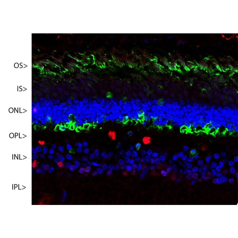
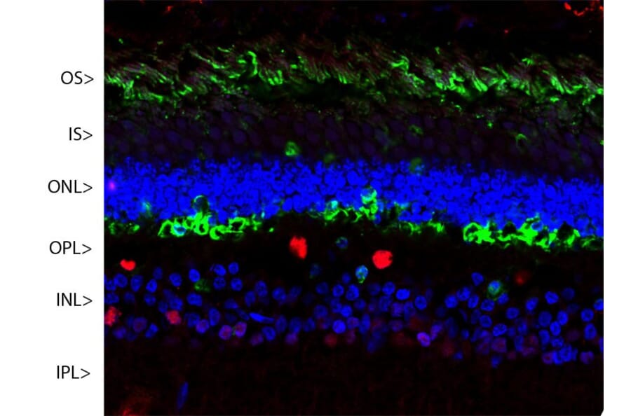
Confocal image of a pig retina stained with Anti-Arrestin-1 Antibody (green). Visual arrestin is most abundant in the outer segments (OS) and inner surface of the outer nuclear layer (ONL), and can be used to identify components of rod photoreceptor cells. (Cone photoreceptors have a different arrestin isotype). Other retinal layers are inner segments (IS), outer plexiform layer (OPL), inner nuclear layer (INL) and inner plexiform layer (IPL). The red stain is from Anti-Fox2 Antibody which stains nuclei of horizontal neurons and some other neurons in the INL and IPL. Nuclear DNA was revealed with DAPI (blue).