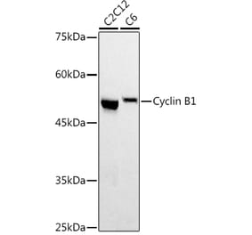
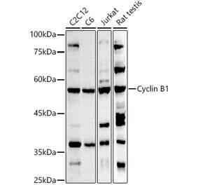
Figure 1: Western Blot - Anti-Cyclin B1 Antibody [ARC2614] (A309196)
Western blot analysis of Jurkat, using Anti-Cyclin B1 Antibody [ARC2614] (A309196) at 1:1,000 dilution. The secondary antibody was Goat Anti-Rabbit IgG H&L Antibody (HRP) at 1:10,000 dilution. Lysates/proteins were present at 25µg per lane. The blocking buffer used was 3% non-fat dry milk in TBST. Detection was with a ECL Basic Kit. Exposure time: 30s.
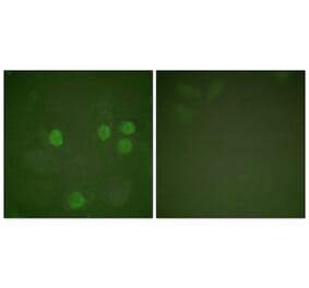
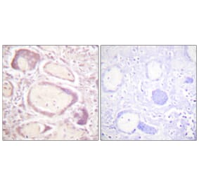
Immunohistochemistry analysis of paraffin-embedded human colon carcinoma tissue using Anti-Cyclin B1 Antibody [ARC2614] (A309196) at a dilution of 1:100 (40x lens). Perform high pressure antigen retrieval with 10 mM citrate buffer pH 6.0 before commencing with IHC staining protocol.
Figure 3: Western Blot - Anti-Cyclin B1 Antibody [ARC2614] (A309196)
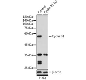
Immunoprecipitation analysis of 300µg extracts of HeLa cells using 3µg of Anti-Cyclin B1 Antibody [ARC2614] (A309196). This Western blot was performed on the immunoprecipitate using Anti-Cyclin B1 Antibody [ARC2614] (A309196) at a dilution of 1:1000.
Alternative Produkte zum Anti-Cyclin B1 Antikörper [ARC2614] (A309196)


![Western Blot - Anti-Cyclin B1 Antibody [ARC2614] (A309196) - Antibodies.com](https://cdn.antibodies.com/image/catalog/309/A309196_1.jpg?profile=product_top)
![Immunohistochemistry - Anti-Cyclin B1 Antibody [ARC2614] (A309196) - Antibodies.com](https://cdn.antibodies.com/image/catalog/309/A309196_2.jpg?profile=product_top)
![Western Blot - Anti-Cyclin B1 Antibody [ARC2614] (A309196) - Antibodies.com](https://cdn.antibodies.com/image/catalog/309/A309196_3.jpg?profile=product_top)
![Western Blot - Anti-Cyclin B1 Antibody [ARC2614] (A309196) - Antibodies.com](https://cdn.antibodies.com/image/catalog/309/A309196_1.jpg?profile=product_top_thumb)
![Immunohistochemistry - Anti-Cyclin B1 Antibody [ARC2614] (A309196) - Antibodies.com](https://cdn.antibodies.com/image/catalog/309/A309196_2.jpg?profile=product_top_thumb)
![Western Blot - Anti-Cyclin B1 Antibody [ARC2614] (A309196) - Antibodies.com](https://cdn.antibodies.com/image/catalog/309/A309196_3.jpg?profile=product_top_thumb)
![Western Blot - Anti-Cyclin B1 Antibody [ARC2614] (A309196) - Antibodies.com](https://cdn.antibodies.com/image/catalog/309/A309196_1.jpg?profile=product_image)
![Immunohistochemistry - Anti-Cyclin B1 Antibody [ARC2614] (A309196) - Antibodies.com](https://cdn.antibodies.com/image/catalog/309/A309196_2.jpg?profile=product_image)
![Western Blot - Anti-Cyclin B1 Antibody [ARC2614] (A309196) - Antibodies.com](https://cdn.antibodies.com/image/catalog/309/A309196_3.jpg?profile=product_image)
![Immunohistochemistry - Anti-Cyclin B1 Antibody [CCNB1/1098] - BSA and Azide free (A253640) - Antibodies.com](https://cdn.antibodies.com/image/catalog/253/A253640_1.jpg?profile=product_alternative)
![Immunohistochemistry - Anti-Cyclin B1 Antibody [CCNB1/1098] (A250460) - Antibodies.com](https://cdn.antibodies.com/image/catalog/250/A250460_1.jpg?profile=product_alternative)
![SDS-PAGE - Anti-Cyclin B1 Antibody [V92.1] (A250459) - Antibodies.com](https://cdn.antibodies.com/image/catalog/250/A250459_1.jpg?profile=product_alternative)
![SDS-PAGE - Anti-Cyclin B1 Antibody [V92.1] - BSA and Azide free (A253639) - Antibodies.com](https://cdn.antibodies.com/image/catalog/253/A253639_1.jpg?profile=product_alternative)
![Immunohistochemistry - Anti-Cyclin B1 Antibody [SPM619] (A250461) - Antibodies.com](https://cdn.antibodies.com/image/catalog/250/A250461_1.jpg?profile=product_alternative)
![Immunohistochemistry - Anti-Cyclin B1 Antibody [SPM619] - BSA and Azide free (A253641) - Antibodies.com](https://cdn.antibodies.com/image/catalog/253/A253641_1.jpg?profile=product_alternative)