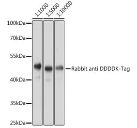
Figure 1: Western Blot - Anti-DDDDK Tag Antibody [ARC5111-01] (A305669)
Western blot analysis of 293T, 293T transfected with GSK3B Protein, 293T transfected with BCAT2-N Protein and 293T transfected with BCAT2-C Protein, using Anti-DDDDK Tag Antibody [ARC5111-01] (A305669) at 1:10,000 dilution. Lysates/proteins were present at 25µg per lane. The blocking buffer used was 3% non-fat dry milk in TBST. Detection was with a ECL Basic Kit. Exposure time: 10s.
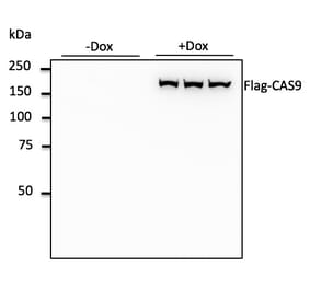
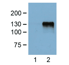
Figure 2: Western Blot - Anti-DDDDK Tag Antibody [ARC5111-01] (A305669)
Western blot analysis of 293F transfected with COPB2-N Protein and 293F transfected with COPB2-C Protein, using Anti-DDDDK Tag Antibody [ARC5111-01] (A305669) at 1:10,000 dilution. Lysates/proteins were present at 25µg per lane. The blocking buffer used was 3% non-fat dry milk in TBST. Detection was with a ECL Basic Kit. Exposure time: 30s.
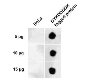
Figure 3: Western Blot - Anti-DDDDK Tag Antibody [ARC5111-01] (A305669)
Western blot analysis of 293T, 293T transfected with IRF1 Protein using Anti-DDDDK Tag Antibody [ARC5111-01] (A305669) at 1:10,000 dilution. Lysates/proteins were present at 25µg per lane. The blocking buffer used was 3% non-fat dry milk in TBST. Detection was with a ECL Basic Kit. Exposure time: 30s.
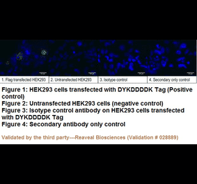
Figure 4: Immunofluorescence - Anti-DDDDK Tag Antibody [ARC5111-01] (A305669)
Immunofluorescence analysis of 293T-Flag-C and 293T-Flag-N and 293T cells using Anti-DDDDK Tag Antibody [ARC5111-01] (A305669) at a dilution of 1:100 (40x lens). DAPI was used to stain the cell nuclei (blue).
Figure 5: Western Blot - Anti-DDDDK Tag Antibody [ARC5111-01] (A305669)
Immunoprecipitation analysis of 300µg extract cell lysate from 293T cells transfected with SERPINB1 expression vector containing a DDDDK-Tag using 3µg of Anti-DDDDK Tag Antibody [ARC5111-01] (A305669). This Western blot was performed on the immunoprecipitate using Anti-DDDDK Tag Antibody [ARC5111-01] (A305669) at a dilution of 1:400.
Figure 6: Western Blot - Anti-DDDDK Tag Antibody [ARC5111-01] (A305669)
Immunoprecipitation analysis of 300µg extract cell lysate from 293T cells transfected with GSK3B expression vector containing a DDDDK-Tag using 3µg of Anti-DDDDK Tag Antibody [ARC5111-01] (A305669). This Western blot was performed on the immunoprecipitate using Anti-DDDDK Tag Antibody [ARC5111-01] (A305669) at a dilution of 1:400.
Figure 7: Flow Cytometry - Anti-DDDDK Tag Antibody [ARC5111-01] (A305669)
Flow cytometry analysis of 293T(Transfection) cells, stained with Rabbit IgG isotype control (2.5 µg/ml, blue line) or Anti-DDDDK Tag Antibody [ARC5111-01] (A305669), (2.5 µg/ml orange line), followed by FITC conjugated goat anti-rabbit polyclonal antibody (1:200 dilution) staining. Non-fluorescently stained 293T(Transfection) cells, used as blank control (red line).
Figure 8: Flow Cytometry - Anti-DDDDK Tag Antibody [ARC5111-01] (A305669)
Flow cytometry analysis of 293T cells, stained with Rabbit IgG isotype control (2.5 µg/ml, blue line) or Anti-DDDDK Tag Antibody [ARC5111-01] (A305669), (2.5 µg/ml orange line), followed by FITC conjugated goat anti-rabbit polyclonal antibody (1:200 dilution). Non-fluorescently stained 293T cells, used as blank control (red line).


![Western Blot - Anti-DDDDK Tag Antibody [ARC5111-01] (A305669) - Antibodies.com](https://cdn.antibodies.com/image/catalog/305/A305669_1.jpg?profile=product_top)
![Western Blot - Anti-DDDDK Tag Antibody [ARC5111-01] (A305669) - Antibodies.com](https://cdn.antibodies.com/image/catalog/305/A305669_2.jpg?profile=product_top)
![Western Blot - Anti-DDDDK Tag Antibody [ARC5111-01] (A305669) - Antibodies.com](https://cdn.antibodies.com/image/catalog/305/A305669_3.jpg?profile=product_top)
![Immunofluorescence - Anti-DDDDK Tag Antibody [ARC5111-01] (A305669) - Antibodies.com](https://cdn.antibodies.com/image/catalog/305/A305669_4.jpg?profile=product_top)
![Western Blot - Anti-DDDDK Tag Antibody [ARC5111-01] (A305669) - Antibodies.com](https://cdn.antibodies.com/image/catalog/305/A305669_5.jpg?profile=product_top)
![Western Blot - Anti-DDDDK Tag Antibody [ARC5111-01] (A305669) - Antibodies.com](https://cdn.antibodies.com/image/catalog/305/A305669_6.jpg?profile=product_top)
![Flow Cytometry - Anti-DDDDK Tag Antibody [ARC5111-01] (A305669) - Antibodies.com](https://cdn.antibodies.com/image/catalog/305/A305669_7.jpg?profile=product_top)
![Flow Cytometry - Anti-DDDDK Tag Antibody [ARC5111-01] (A305669) - Antibodies.com](https://cdn.antibodies.com/image/catalog/305/A305669_8.jpg?profile=product_top)
![Western Blot - Anti-DDDDK Tag Antibody [ARC5111-01] (A305669) - Antibodies.com](https://cdn.antibodies.com/image/catalog/305/A305669_1.jpg?profile=product_top_thumb)
![Western Blot - Anti-DDDDK Tag Antibody [ARC5111-01] (A305669) - Antibodies.com](https://cdn.antibodies.com/image/catalog/305/A305669_2.jpg?profile=product_top_thumb)
![Western Blot - Anti-DDDDK Tag Antibody [ARC5111-01] (A305669) - Antibodies.com](https://cdn.antibodies.com/image/catalog/305/A305669_3.jpg?profile=product_top_thumb)
![Immunofluorescence - Anti-DDDDK Tag Antibody [ARC5111-01] (A305669) - Antibodies.com](https://cdn.antibodies.com/image/catalog/305/A305669_4.jpg?profile=product_top_thumb)
![Western Blot - Anti-DDDDK Tag Antibody [ARC5111-01] (A305669) - Antibodies.com](https://cdn.antibodies.com/image/catalog/305/A305669_5.jpg?profile=product_top_thumb)
![Western Blot - Anti-DDDDK Tag Antibody [ARC5111-01] (A305669) - Antibodies.com](https://cdn.antibodies.com/image/catalog/305/A305669_6.jpg?profile=product_top_thumb)
![Flow Cytometry - Anti-DDDDK Tag Antibody [ARC5111-01] (A305669) - Antibodies.com](https://cdn.antibodies.com/image/catalog/305/A305669_7.jpg?profile=product_top_thumb)
![Immunocytochemistry - Anti-DDDDK Tag Antibody [F-tag-01] (A85906) - Antibodies.com](https://cdn.antibodies.com/image/catalog/85/A85906_301.jpg?profile=product_alternative)
![Western Blot - Anti-DDDDK Tag Antibody [ARC5111-01] (A305669) - Antibodies.com](https://cdn.antibodies.com/image/catalog/305/A305669_1.jpg?profile=product_image)
![Western Blot - Anti-DDDDK Tag Antibody [ARC5111-01] (A305669) - Antibodies.com](https://cdn.antibodies.com/image/catalog/305/A305669_2.jpg?profile=product_image)
![Western Blot - Anti-DDDDK Tag Antibody [ARC5111-01] (A305669) - Antibodies.com](https://cdn.antibodies.com/image/catalog/305/A305669_3.jpg?profile=product_image)
![Immunofluorescence - Anti-DDDDK Tag Antibody [ARC5111-01] (A305669) - Antibodies.com](https://cdn.antibodies.com/image/catalog/305/A305669_4.jpg?profile=product_image)
![Western Blot - Anti-DDDDK Tag Antibody [ARC5111-01] (A305669) - Antibodies.com](https://cdn.antibodies.com/image/catalog/305/A305669_5.jpg?profile=product_image)
![Western Blot - Anti-DDDDK Tag Antibody [ARC5111-01] (A305669) - Antibodies.com](https://cdn.antibodies.com/image/catalog/305/A305669_6.jpg?profile=product_image)
![Flow Cytometry - Anti-DDDDK Tag Antibody [ARC5111-01] (A305669) - Antibodies.com](https://cdn.antibodies.com/image/catalog/305/A305669_7.jpg?profile=product_image)
![Flow Cytometry - Anti-DDDDK Tag Antibody [ARC5111-01] (A305669) - Antibodies.com](https://cdn.antibodies.com/image/catalog/305/A305669_8.jpg?profile=product_image)