Synthetic peptide derived from human JAK1 (amino acids 988-1037).
Wirt
Rabbit
Klonalität
Polyclonal
Isotyp
IgG
Konjugat
Unconjugated
Reinigung
Purified from rabbit serum by antigen affinity chromatography using the immunizing peptide.
Molekulargewicht
131kDa
Produktform
Liquid
Formulierung
Supplied in Phosphate Buffered Saline (without Mg2+ and Ca2+), pH 7.4, with 150mM NaCl, 0.02% Sodium Azide, and 50% Glycerol.
Lagerung
Shipped at 4°C. Upon delivery aliquot and store at -20°C. Avoid freeze / thaw cycles.
Synonyme
JAK 1, JAK 1A, JAK 1B, JAK-1, JAK1A, JAK1B, JAK1_HUMAN, Janus kinase 1, Janus kinase 1 (a protein tyrosine kinase), JTK3, Tyrosine protein kinase JAK 1, Tyrosine protein kinase JAK1, Tyrosine-protein kinase JAK1
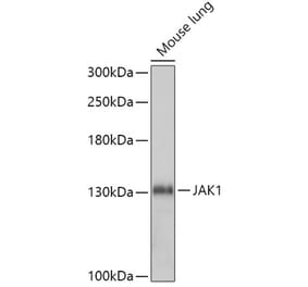
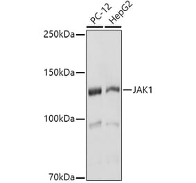
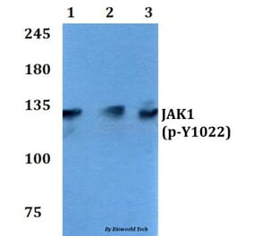
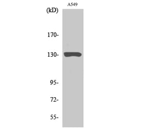
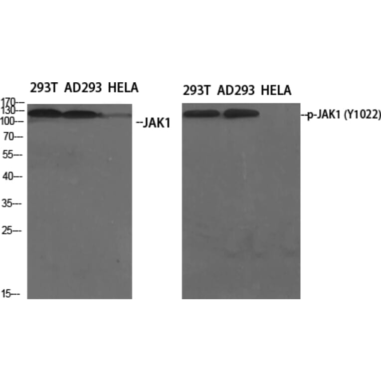
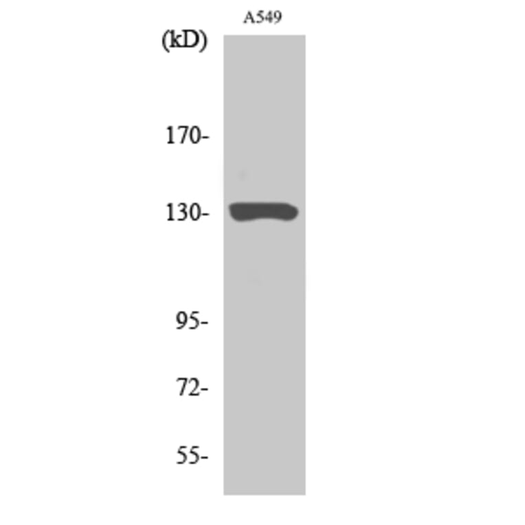
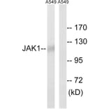
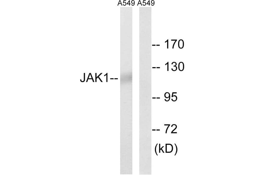
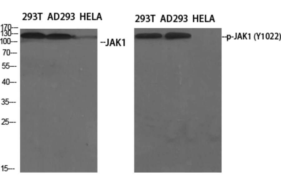
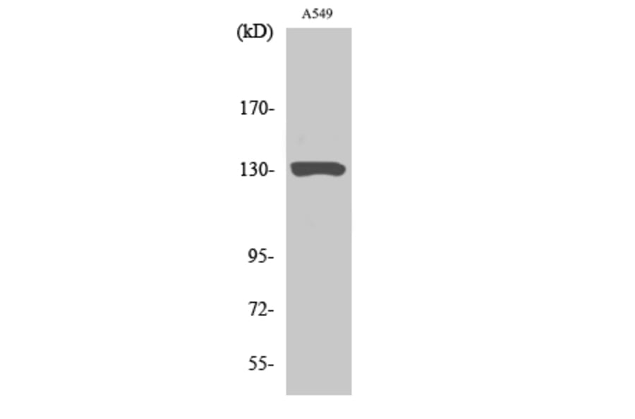
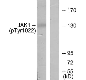
Figure 1: Western Blot - Anti-JAK1 Antibody (A96173)
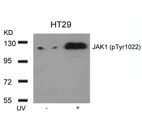
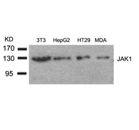
Western blot analysis of lysates from A549 using Anti-JAK1 Antibody. The right hand lane represents a negative control, where the antibody is blocked by the immunising peptide.
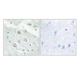
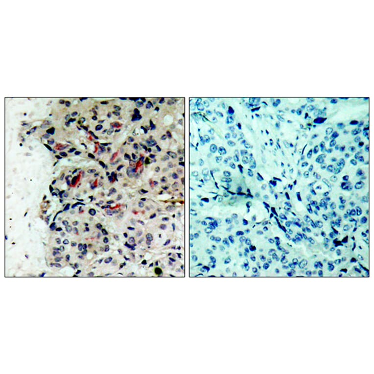
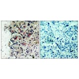
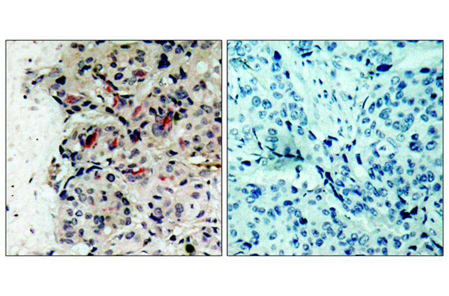
Immunohistochemical analysis of paraffin-embedded human breast carcinoma tissue using Anti-JAK1 Antibody. The right hand panel represents a negative control, where the antibody was pre-incubated with the immunising peptide.
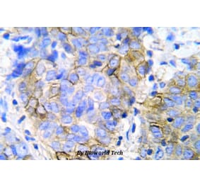
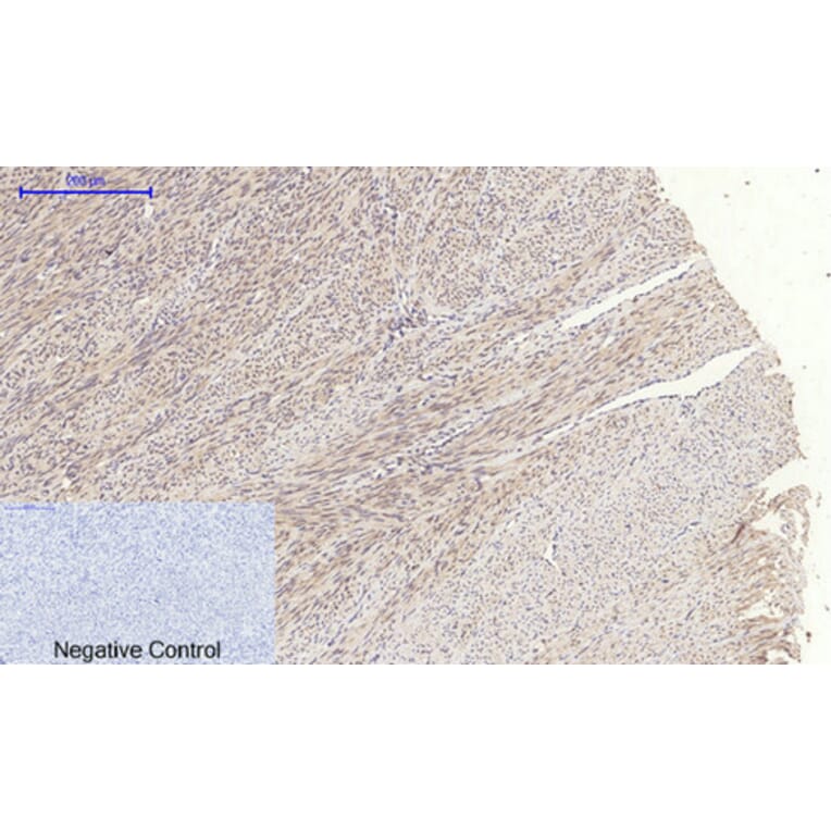
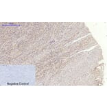
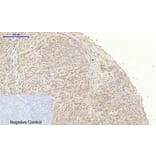
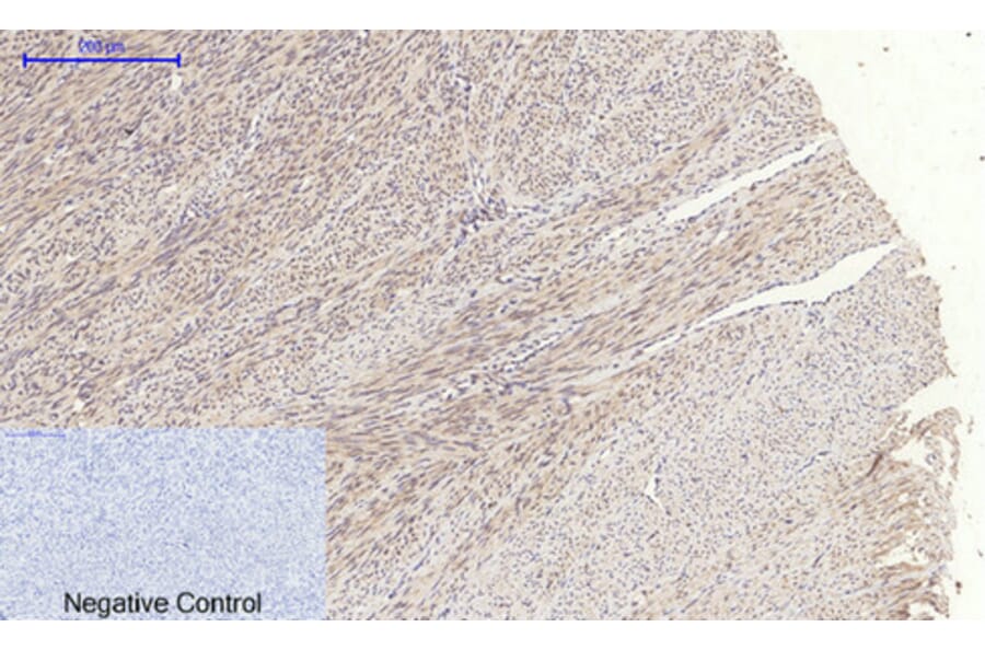
Immunohistochemical analysis of paraffin-embedded human uterus tissue using Anti-JAK1 Antibody at 1:200 (4°C overnight). Negative control was secondary antibody only.
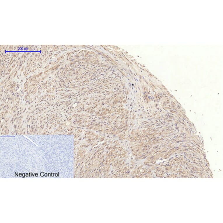
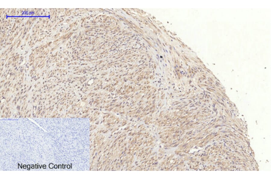
Immunohistochemical analysis of paraffin-embedded human uterus cancer tissue using Anti-JAK1 Antibody at 1:200 (4°C overnight). Negative control was secondary antibody only.
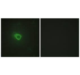
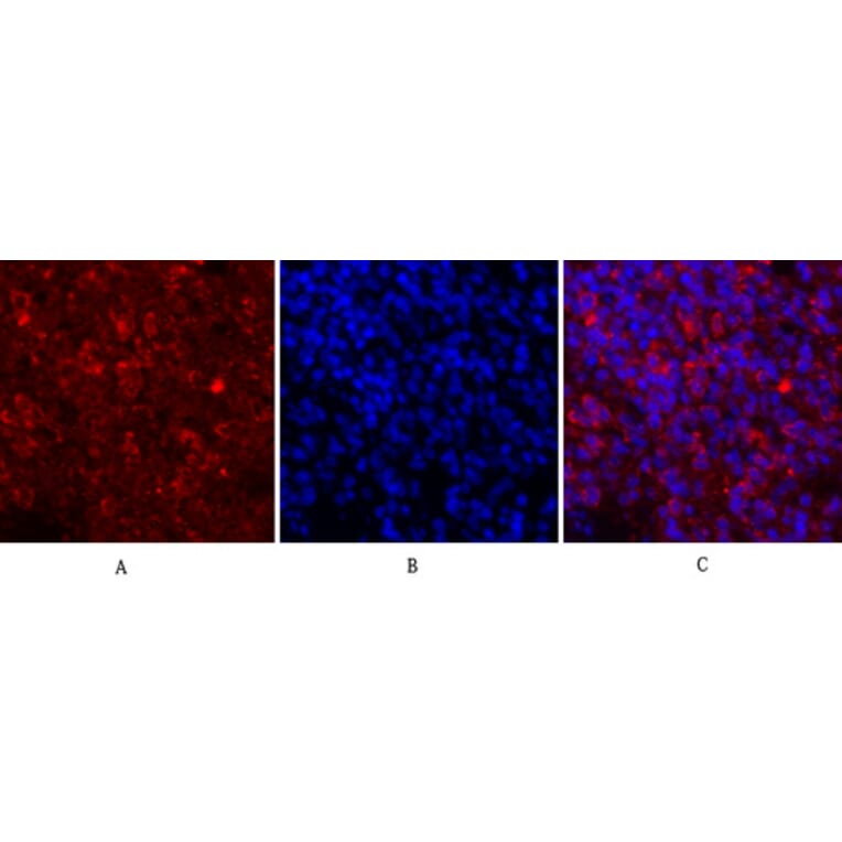
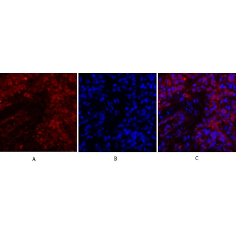
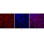
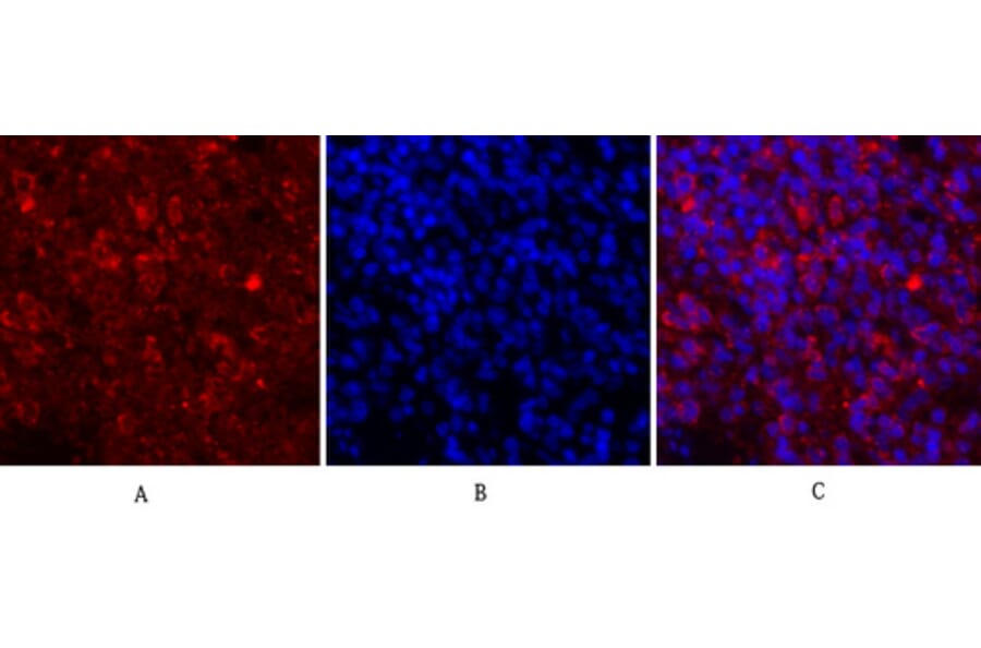
Immunofluorescence analysis of rat spleen tissue using Anti-JAK1 Antibody (red) at 1:200 (4°C overnight). Cy3 labelled secondary antibody was used at 1:300 (RT 50min). Panel A: Target. Panel B: DAPI. Panel C: Merge.
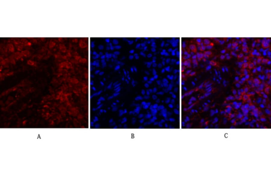
Immunofluorescence analysis of rat lung tissue using Anti-JAK1 Antibody (red) at 1:200 (4°C overnight). Cy3 labelled secondary antibody was used at 1:300 (RT 50min). Panel A: Target. Panel B: DAPI. Panel C: Merge.


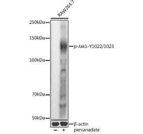
![Western Blot - Anti-JAK1 Antibody [ARC0434] (A80915) - Antibodies.com](https://cdn.antibodies.com/image/catalog/80/A80915_1.jpg?profile=product_alternative)