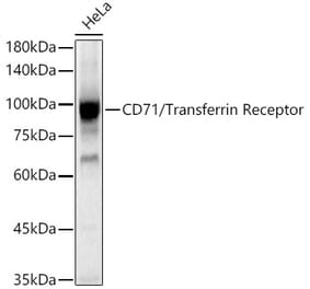
Figure 1: Western Blot - Anti-Transferrin Receptor Antibody [ARC54396] (A308474)
Western blot analysis of HeLa, using Anti-Transferrin Receptor Antibody [ARC54396] (A308474) at 1:80000 dilution. The secondary antibody was Goat Anti-Rabbit IgG H&L Antibody (HRP) at 1:10,000 dilution. Lysates/proteins were present at 25µg per lane. The blocking buffer used was 3% non-fat dry milk in TBST. Detection was with a ECL Basic Kit. Exposure time: 5s.
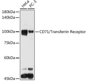
Figure 2: Western Blot - Anti-Transferrin Receptor Antibody [ARC54396] (A308474)
Western blot analysis of various lysates, using Anti-Transferrin Receptor Antibody [ARC54396] (A308474) at 1:80000 dilution. The secondary antibody was Goat Anti-Rabbit IgG H&L Antibody (HRP) at 1:10,000 dilution. Lysates/proteins were present at 25µg per lane. The blocking buffer used was 3% non-fat dry milk in TBST. Detection was with a ECL Basic Kit. Exposure time: 60s.
Immunohistochemistry analysis of paraffin-embedded mouse liver using Anti-Transferrin Receptor Antibody [ARC54396] (A308474) at a dilution of 1:100(40x lens). Perform high pressure antigen retrieval with 10 mM citrate buffer pH 6.0 before commencing with IHC staining protocol.
Confocal imaging of HeLa cells using Anti-Transferrin Receptor Antibody [ARC54396] (A308474), at a dilution of 1:200, (red). The cells were counterstained with Anti-CD44 Antibody, at a dilution of 1:200, (green). DAPI was used for nuclear staining (Blue). Objective: 60x.
Alternative Produkte zum Anti-Transferrin Receptor Antikörper [ARC54396] (A308474)


![Western Blot - Anti-Transferrin Receptor Antibody [ARC54396] (A308474) - Antibodies.com](https://cdn.antibodies.com/image/catalog/308/A308474_1.jpg?profile=product_top)
![Western Blot - Anti-Transferrin Receptor Antibody [ARC54396] (A308474) - Antibodies.com](https://cdn.antibodies.com/image/catalog/308/A308474_2.jpg?profile=product_top)
![Immunohistochemistry - Anti-Transferrin Receptor Antibody [ARC54396] (A308474) - Antibodies.com](https://cdn.antibodies.com/image/catalog/308/A308474_3.jpg?profile=product_top)
![Immunofluorescence - Anti-Transferrin Receptor Antibody [ARC54396] (A308474) - Antibodies.com](https://cdn.antibodies.com/image/catalog/308/A308474_4.jpg?profile=product_top)
![Western Blot - Anti-Transferrin Receptor Antibody [ARC54396] (A308474) - Antibodies.com](https://cdn.antibodies.com/image/catalog/308/A308474_1.jpg?profile=product_top_thumb)
![Western Blot - Anti-Transferrin Receptor Antibody [ARC54396] (A308474) - Antibodies.com](https://cdn.antibodies.com/image/catalog/308/A308474_2.jpg?profile=product_top_thumb)
![Immunohistochemistry - Anti-Transferrin Receptor Antibody [ARC54396] (A308474) - Antibodies.com](https://cdn.antibodies.com/image/catalog/308/A308474_3.jpg?profile=product_top_thumb)
![Immunofluorescence - Anti-Transferrin Receptor Antibody [ARC54396] (A308474) - Antibodies.com](https://cdn.antibodies.com/image/catalog/308/A308474_4.jpg?profile=product_top_thumb)
![Western Blot - Anti-Transferrin Receptor Antibody [ARC54396] (A308474) - Antibodies.com](https://cdn.antibodies.com/image/catalog/308/A308474_1.jpg?profile=product_image)
![Western Blot - Anti-Transferrin Receptor Antibody [ARC54396] (A308474) - Antibodies.com](https://cdn.antibodies.com/image/catalog/308/A308474_2.jpg?profile=product_image)
![Immunohistochemistry - Anti-Transferrin Receptor Antibody [ARC54396] (A308474) - Antibodies.com](https://cdn.antibodies.com/image/catalog/308/A308474_3.jpg?profile=product_image)
![Immunofluorescence - Anti-Transferrin Receptor Antibody [ARC54396] (A308474) - Antibodies.com](https://cdn.antibodies.com/image/catalog/308/A308474_4.jpg?profile=product_image)
![Flow Cytometry - Anti-CD71 Antibody [MEM-189] (A85951) - Antibodies.com](https://cdn.antibodies.com/image/catalog/85/A85952_329.jpg?profile=product_alternative)
![Immunocytochemistry - Anti-CD71 Antibody [MEM-75] (A85652) - Antibodies.com](https://cdn.antibodies.com/image/catalog/85/A85654_132.jpg?profile=product_alternative)
![SDS-PAGE - Anti-Transferrin Receptor Antibody [Jr-141] - Low endotoxin, Azide free (A324276) - Antibodies.com](https://cdn.antibodies.com/image/catalog/324/A324276_1.jpg?profile=product_alternative)
![Immunofluorescence - Anti-Transferrin Receptor Antibody [DF1513] (A250092) - Antibodies.com](https://cdn.antibodies.com/image/catalog/250/A250092_1.jpg?profile=product_alternative)
![Immunofluorescence - Anti-Transferrin Receptor Antibody [DF1513] - BSA and Azide free (A253272) - Antibodies.com](https://cdn.antibodies.com/image/catalog/253/A253272_1.jpg?profile=product_alternative)
![Immunohistochemistry - Anti-Transferrin Receptor Antibody [TFRC/1817] (A250096) - Antibodies.com](https://cdn.antibodies.com/image/catalog/250/A250097_1.jpg?profile=product_alternative)
![Flow Cytometry - Anti-Transferrin Receptor Antibody [66IG10] (A250093) - Antibodies.com](https://cdn.antibodies.com/image/catalog/250/A250093_1.jpg?profile=product_alternative)
![Flow Cytometry - Anti-Transferrin Receptor Antibody [66IG10] - BSA and Azide free (A253273) - Antibodies.com](https://cdn.antibodies.com/image/catalog/253/A253273_1.jpg?profile=product_alternative)