ELISpot Kits
Michaela Buerdsell, PhD and Kay Shigemori, PhD | 15th September 2026
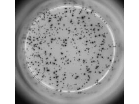
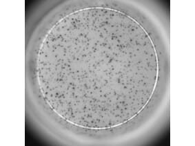
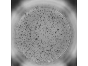
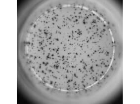
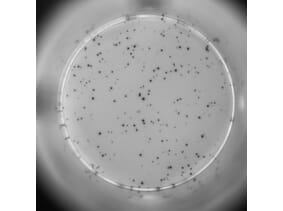
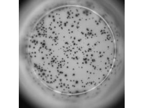
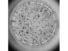
Enzyme-Linked ImmunoSpot (ELISpot) is a highly sensitive immunoassay used to detect and quantify individual immune cells that secrete analytes such as cytokines and antibodies at a single-cell level. Each spot represents a single secreting cell, while the size of the spot and intensity can provide information on the magnitude of the secretion. ELISpot assays are widely used in both research and clinical settings such as for vaccine-induced immunity, monitor immunotherapy responses, and study autoimmune diseases. Our catalogue includes 50 validated ELISpot assay kits, delivering exceptional senstivity and reproducibility.
Table of Contents
What is ELISpot
Enzyme-linked ImmunoSpot (ELISpot) is a plate based immunoassay used to identify secreted products, normally cytokines, from an immune cell of choice from the adaptive or innate immune systems.1 Secreted analytes will form small spots composed of the enzyme-substrate precipitates, which can be counted on an ELISpot analyzer or by manually counting the spots using a microscope.2 Each spot corresponds to a single secreting cell, as the antibodies rapidly bind to the analyte surrounding that cell.3 Measuring the frequency of the spots can provide information on the number of secreting cells whilst the size of the spot can correlate to secretion magnitude.4
The ability of ELISpot assays to sensitively enumerate cytokine, antibody, and other secretions from individual immune cells has both in ex vivo and in vitro settings has led to the widespread use in across immunological research and clinical applications. For example, ELISpot assays were used to identify clinically relevant immune biomarkers in chronic hepatitis, and monitor immune responses following treatment to patients with sepsis, helping characterize immune profiles and the effect of immune-modulatory drugs.5,6 In clinical settings, ELISpot assays have been used to monitor antigen-specific immune responses following vaccination and to evaluate immune activity during cancer immunotherapy and other therapeutic interventions.1,4
How to Perform an ELISpot Assay
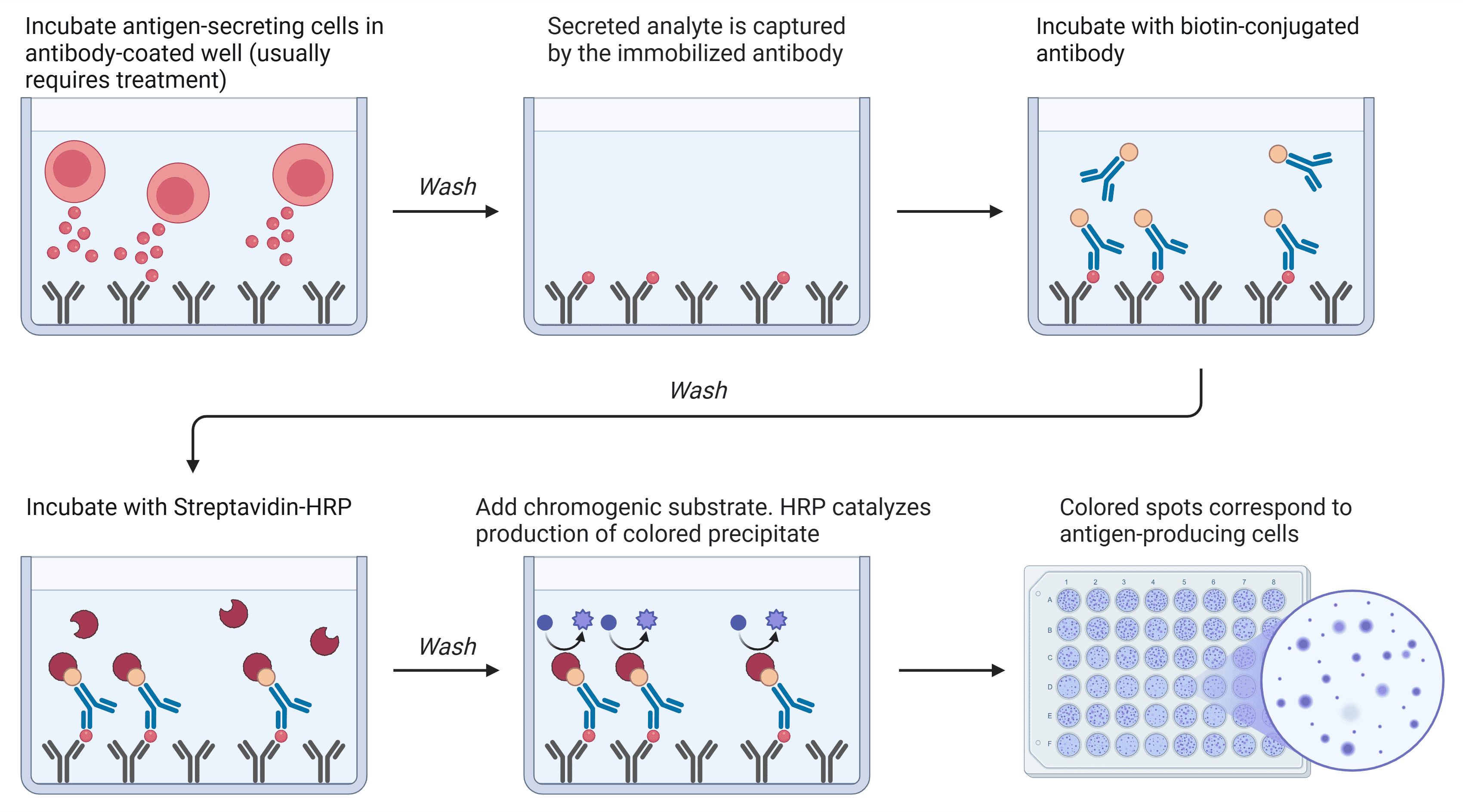
Most ELISpot assays follow the same core principle: immobilize capture antibody, incubate cells so analyte is captured locally, detect captured analyte using labelled detection reagents and an insoluble reporter that forms spots at secretion sites (Figure 1).4,7
Diagram created with BioRender.
Step 1: Coat the plate with capture antibodies
- Prepare membrane plate (PVDF/nitrocellulose; activate PVDF if required)
- Coat with capture antibody (often overnight at 4 °C)
- Block non-specific binding sites
Step 2: Add cells & stimulating agents; activated cells secrete cytokines
- Add cells, stimulating agent, and controls
- Incubate (commonly 16–24 h for many cytokines)
Step 3: Remove cells and add an enzyme-labelled detection antibody
- Wash thoroughly to remove cells
- Add biotinylated detection antibody and streptavidin-enzyme (AP/HRP)
- Wash thoroughly to remove excess conjugated detection antibody
Step 4: Add substrate to visualize spots
- Add precipitating substrate
- Develop spots
Step 5: Spot detection and quantification
- Dry and scan with ELISpot plate reader on recommended settings (or count manually using a microscope)
- Quantify the number of spots (SPU per 106 cells)
Workflow reference: Cox et al. (2006) Janetzki et al. (2015).
Why ELISpot? The Advantages
ELISpot is widely used because it combines high sensitivity with functional relevance:
- Single Cell Resolution: detects rare antigen-specific secreting cells at very low frequencies.4
- Very high sensitivity and low background: local capture improves signal discrimination when optimized.7
- Quantitative and comparable: harmonized layouts and automated evaluation improved reproducibility.
- Direct functional readout: measures secreted proteins rather than intracellular accumulation.4
- Streamlined workflows: assay ready kits (pre-coated PVDF with capture antibody, validated antibody pairs, stimulants) reduce setup time and variability.
When to Use ELISpot
ELISpot and related FluoroSpot and Dual color assays are broadly used in immunology research and translational settings to quantify antigen‑specific, cytokine‑secreting cells.4 ELISpot quantifies the number of cells actively secreting a defined analyte (typically a cytokine or antibody), making it well-suited for measuring responder frequency, for example, quantifying IFN-γ-secreting T cells after peptide stimulation or antibody-secreting B cells following immunization.3,8 ELISpot is particularly advantageous when the expected response is rare, the sample input is limited, or robust standardization is required across experiments or sites.
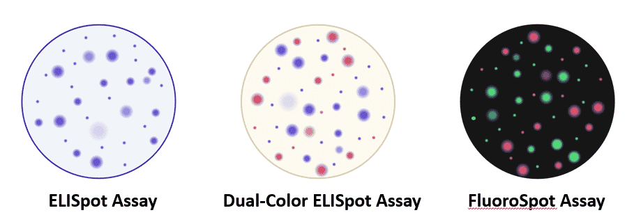
A dual-color ELISpot is a modification of the ELISpot assay that uses different enzyme-substrate reactions to generate distinct colors, enabling the simultaneous detection of two or three types of secreting cells.9 The resulting enzyme-substrate precipitates form different colored spots that correspond to a specific type of secreting immune cells. One such example used dual-color ELISpot assays to simultaneously detect IL-2 and IFN- γ secreting cells to characterize the immune response to HIV antigens.10 This type of assay still utilizes a colorimetric system, however an alternative adaption known as FluoroSpot assay uses fluorescence-based detection to identify multiple analytes within the same well.2 The sensitivity of FluoroSpot assays have been shown to be comparable to ELISpot assays but using different fluorescent markers allows for the detection of up to three cytokines in the same well allowing for broader characterization of immune responses.11
Diagram created with BioRender.
ELISpot vs ELISA vs Flow Cytometry
ELISpot is best viewed as a functional frequency assay, whereas ELISA measures bulk analyte concentration and flow cytometry (e.g., intracellular cytokine staining) is used for cellular phenotyping and multiplexed readouts. These methods are complementary and should be chosen depending on whether you are assessing analyte concentration, frequency of secreting cells, or cellular phenotype.4,7
| Feature | ELISpot | ELISA | Flow Cytometry |
| Primary Readout | Frequency of secreting cells (SFU per unit of cells) | Bulk final concentration (pg/mL) | Cell phenotype (% positive/negative cells and/or mean) |
| Sensitivity for Rare Responders | Very High | Depends; no frequency information | Moderate - High |
| Single Cell Resolution | Yes | No | Yes |
| Multiplexing | Dual-Color ELISpot/FluoroSpot | High (bulking multiplex formats) | Very High |
| Sample | Secreting immune cells | Lysed cells or tissue; Biological fluids (plasma, urine) | Often bulk cells |
| Best Suited For | Responder frequency screening | Soluble analyte total quantification | Subset analysis & polyfunctionality |
Applications for ELISpot Assays
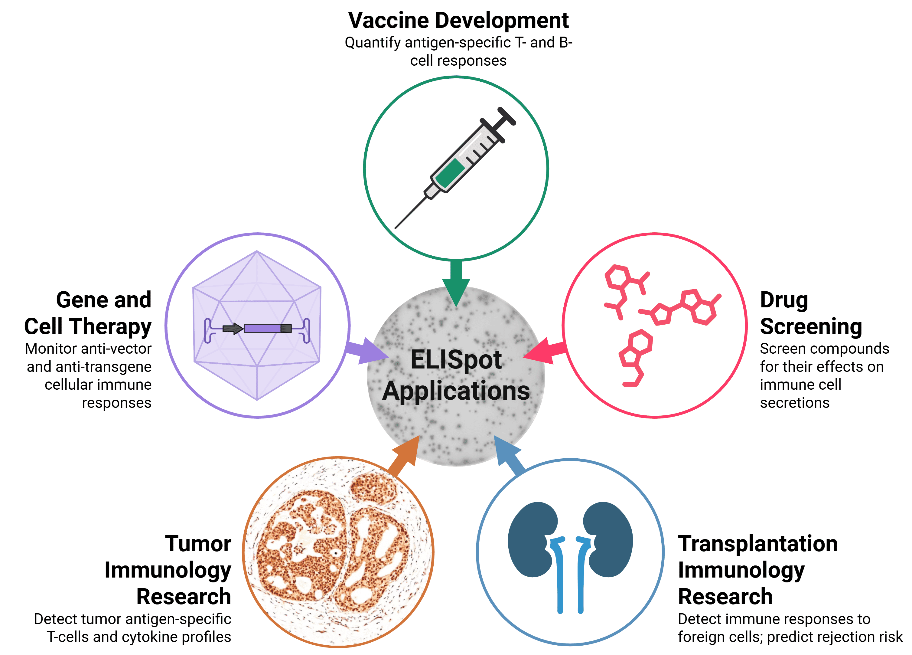
ELISpot assays are used widely in basic, traditional, and clinical research including:
- Vaccines & Infectious Disease: Tracking vaccine or antigen-induced T cell immunity in influenza, HPV, or HIV.13-15
- Cancer Immunotherapy & Cancer Vaccines Monitoring immune responses during cancer treatment to evaluate the efficacy of the immunotherapeutic intervention and correlate to clinical outcomes.3
- Cytotoxicity Profiling Granzyme B/Perforin secretion for monitoring cell-mediated cytotoxicity.16
- Autoimmunity & Allergy Studying hypersensitivity and autoimmunity via IFN- γ, granzyme B, IL-4, IL-5, or IL-13 cytokine response profiling.17
- Clinical Diagnosis: ELISpot-based workflows used in a real-world clinical setting, such as for tuberculosis diagnostics and early diagnostics of tuberculosis meningoencephalitis.18,19
- B cell Immunity/ASC Enumeration: Measuring B cell activity and B cell memory formation by measuring IgG/IgA secretion and antigen-specific antibody secretion.8
Diagram created with BioRender.
Troubleshooting Guide
Most ELISpot issues fall into four categories: cell quality, stimulation conditions, coating/blocking/washing, or detection/development/reader settings. Begin troubleshooting by inspecting negative controls, positive controls, and replicate agreement. Standardized reader settings and evaluation templates are essential for comparability across plates and studies.4,20,21 Below features a table with common ELISpot problems and some suggestions on overcoming the issue.
| Issue | Likely Cause(s) | Practical Fixes | Prevention |
| High Background | - Insufficient Washing
- Over-development
- High concentration of detection reagents/high affinity
| - Titrate detection reagents
- Increase wash/soak steps
- Shorten substrate incubation time
| - Do not let the plate dry
- Include matched vehicle controls
- Optimize washing volume/repeats
|
| Weak/No Signal | - Low cell viability
- Poor stimulation
- Faulty coating
| - Reduce cell input
- Shorten development time
| - Check cell viability using Trypan Blue (or similar)
- Titrate cell number
- Titrate stimulation
|
| Small/Faint Spots | - Low capture of analyte
- Low concentration of detection antibody
- Under-developemnt of substrate
| - Optimize analyte capture antibody
- Optimize detection antibody concentration
- Extend development time
| - Document development time
- Keep development time consistent
|
| High Variability | - Uneven plating
- Edge effects
| - Mix cells during plating by swirling
- Ensure even pipetting between wells
| - Check for uniform cell dispersion
- Pre-warm reagents and media to incubation temperature
|
| Edge Effects | | - Fill outer wells with buffer/media
- Incubate in a humidified incubator
| - Use a humidified incubator
- Use a plate lid
|
| Hazy Staining | - Precipitated substrate
- Poor washing
| - Use fresh substrate
- Increase washing steps
- Wash for longer
| - Reagent QC
- Optimize wash steps
|
| Too Many Spots | - Stimulus too strong
- Saturation
| - Dilute stimulus
- Dilute cell number
| - Define linear assay range
|
| Non-Specific Activation | - Presence of endotoxin
- DMSO Effects
- Stress
| - Use low-endotoxin reagents
- Rest PBMCs
| - Record pre-analytical variables
- Include a vehicle control
|
Overnight resting of thawed PBMC has been shown to improve IFN-γ ELISpot performance in some workflows.22
Featured Comprehensive ELISpot Kits
Suited for peripheral blood mononuclear cells (PBMCs) in single suspension, capable of cytokine secretion.
Suitable for cytokine secreting peripheral blood mononuclear cells (PBMCs) in single suspension.
Suitable for peripheral blood mononuclear cells (PBMCs) in single suspension capable of cytokine secretion.
Suitable for cytokine secreting peripheral blood mononuclear cells (PBMCs) in single suspension.
Suited to secreting peripheral blood mononuclear cells (PBMCs) in single-cell suspension.
Available for cytokine secreting peripheral blood mononuclear cells (PBMCs) in single-cell suspension.
Peripheral blood mononuclear cells (PBMCs) in single-cell suspension, capable of cytokine secretion.
Suitable for peripheral blood mononuclear cells (PBMCs) in single-cell suspension, capable of cytokine secretion.
Suited for cytokine secreting peripheral blood mononuclear cells (PBMCs) in single cell suspension.
References
Diagrams created with BioRender.com.
- Slota, M., Lim, J.-B., Dang, Y. & Disis, M. L. ELISpot for measuring human immune responses to vaccines. Expert Rev. Vaccines 10, 299–306 (2011).
- Janetzki, S. Automation of the Elispot Technique: Past, Present, and Future. SLAS Technol. 9, 10–15 (2004).
- Fernández Castro, L. R. et al. ELISPOT as a Functional for Biomarker Study in Cancer Immunotherapy: Applications and Future Directions. Int. J. Mol. Sci. 27, 4056 (2026).
- Janetzki, S. Immune monitoring technology primer: the enzyme-linked immunospot (Elispot) and Fluorospot assay. J. Immunother. Cancer 3, 30 (2015).
- Gehring, A. J. et al. Immunological biomarker discovery in cure regimens for chronic hepatitis B virus infection. J. Hepatol. 77, 525–538 (2022).
- Walton, A. H. et al. Determining potential immunomodulatory drug efficacy in sepsis using ELISpot. Sci. Rep. 15, 13464 (2025).
- Cox, J. H., Ferrari, G. & Janetzki, S. Measurement of cytokine release at the single cell level using the ELISPOT assay. Methods 38, 274–282 (2006).
- Stylianou, G., Kirchenbaum, G. A., Lehmann, P. V., Pearce, S. & Todryk, S. Measuring Human Memory B Cells in Autoimmunity Using Enzyme-Linked ImmunoSpot. Biomolecules 15, 643 (2025).
- Czerkinsky, C., Moldoveanu, Z., Mestecky, J., Nilsson, L.-Å. & Ouchterlony, Ö. A novel two colour ELISPOT assay: I. Simultaneous detection of distinct types of antibody-secreting cells. J. Immunol. Methods 115, 31–37 (1988).
- Boulet, S. et al. A Dual Color ELISPOT Method for the Simultaneous Detection of IL-2 and IFN-γ HIV-Specific Immune Responses. J. Immunol. Methods 320, 18–29 (2007).
- Dillenbeck, T., Gelius, E., Fohlstedt, J. & Ahlborg, N. Triple Cytokine FluoroSpot Analysis of Human Antigen-Specific IFN-γ, IL-17A and IL-22 Responses. Cells 3, 1116–1130 (2014).
- Waerlop, G. et al. Harmonization and qualification of an IFN-γ Enzyme-Linked ImmunoSpot assay (ELISPOT) to measure influenza-specific cell-mediated immunity within the FLUCOP consortium. Front. Immunol. 13, 984642 (2022).
- Kenter, G. G. et al. Vaccination against HPV-16 Oncoproteins for Vulvar Intraepithelial Neoplasia. N. Engl. J. Med. 361, 1838–1847 (2009).
- Samri, A. et al. Evaluation of the Interlaboratory Concordance in Quantification of Human Immunodeficiency Virus-Specific T Cells with a Gamma Interferon Enzyme-Linked Immunospot Assay. Clin. Vaccine Immunol. 13, 684–697 (2006).
- Malyguine, A. M., Strobl, S., Dunham, K., Shurin, M. R. & Sayers, T. J. ELISPOT Assay for Monitoring Cytotoxic T Lymphocytes (CTL) Activity in Cancer Vaccine Clinical Trials. Cells 1, 111–126 (2012).
- Porebski, G., Piotrowicz-Wojcik, K. & Spiewak, R. ELISpot assay as a diagnostic tool in drug hypersensitivity reactions. J. Immunol. Methods 495, 113062 (2021).
- Wang, J.-Y. et al. Diagnosis of Tuberculosis by an Enzyme-Linked Immunospot Assay for Interferon-γ. Emerg. Infect. Dis. 13, 553–558 (2007).
- Blume, J., Köstler, J. & Weissert, R. Benefit of ELISpot in early diagnosis of tuberculous meningoencephalitis: Case report and literature review. eNeurologicalSci 1, 51–53 (2015).
- Janetzki, S. et al. Guidelines for the automated evaluation of Elispot assays. Nat. Protoc. 10, 1098–1115 (2015).
- Janetzki, S. et al. Results and harmonization guidelines from two large-scale international Elispot proficiency panels conducted by the Cancer Vaccine Consortium (CVC/SVI). Cancer Immunol. Immunother. CII 57, 303–315 (2007).
- Gazagne, A. et al. A Fluorospot assay to detect single T lymphocytes simultaneously producing multiple cytokines. J. Immunol. Methods 283, 91–98 (2003).