Unconjugated
Rapidly progressive glomerulonephritis is characterized by glomerular necroinflammation and crescent formation. Its treatment includes unspecific and toxic agents; therefore, the identification of novel therapeutic targets is required. The E3-ubiquitin ligase murine double minute (MDM)-2 is a nonredundant element of NF-κB signaling and the negative regulator of tumor suppressor gene TP53-mediated cell cycle arrest and cell death. We hypothesized that the MDM2 would drive crescentic glomerulonephritis by NF-κB-dependent glomerular inflammation and by p53-dependent parietal epithelial cell hyperproliferation. Indeed, the pre-emptive MDM2 blockade by nutlin-3a ameliorated all aspects of crescentic glomerulonephritis. MDM2 inhibition had identical protective effects in Trp53-deficient mice, with the exception of crescent formation, which was not influenced by nutlin-3a treatment. In vitro experiments confirmed the contribution of MDM2 for induction of NF-κB-dependent cytokines in murine glomerular endothelial cells and for p53-dependent parietal epithelial cell proliferation. To evaluate MDM2 blockade as a potential therapeutic intervention in rapidly progressive glomerulonephritis, we treated mice with established glomerulonephritis with nutlin-3a. Delayed onset of nutlin-3a treatment was equally protective as the pre-emptive treatment in abrogating crescentic glomerulonephritis. Together, the pathogenic effects of MDM2 are twofold, that is, p53-independent NF-κB activation increasing intraglomerular inflammation and p53-dependent parietal epithelial cell hyperplasia and crescent formation. We therefore propose MDM2 blockade as a potential novel therapeutic strategy in rapidly progressive glomerulonephritis.
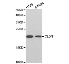
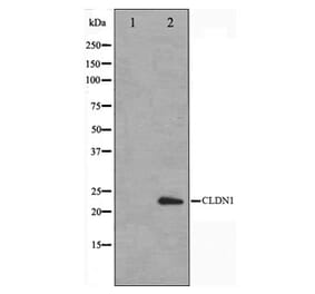
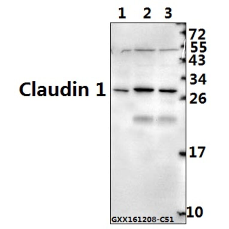
This study investigated the effect of phytic acid (IP6), a potential absorption enhancer of flavonoid components, on tight junction (TJ) integrity in Caco-2 cell monolayers and its possible mechanisms. Transepithelial electrical resistance (TEER) across the monolayers decreased rapidly, and the flux of fluorescein sodium (a paracellular marker) increased after treating with IP6 in a concentration-dependent manner. Confocal microscopy results showed that IP6 produced a concentration-dependent attenuation in the distribution of occludin, ZO-1, and claudin-1. Immunoblot analysis revealed that IP6 could down-regulate the expression level of these TJ proteins, which resulted in the opening of TJ. Additionally, the divalent cations Ca(2+) and Mg(2+) influenced the IP6-induced distribution of occludin, ZO-1, and claudin-1 in different directions, which enhanced barrier function. In conclusion, IP6 can decrease the integrity of Caco-2 cell monolayers by modulating the TJ proteins' localization and down-regulating the expression levels of TJ proteins including claudin-1, occludin, and ZO-1; the reduction effects of divalent cations such as Ca(2+) and Mg(2+) on the regulation of TJ induced by IP6 should be addressed. The present work will offer some useful guidance for the application of IP6 in drug delivery area.


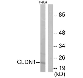
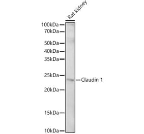
![Western Blot - Anti-Claudin 1 Antibody [ARC54475] (A305830) - Antibodies.com](https://cdn.antibodies.com/image/catalog/305/A305830_1.jpg?profile=product_alternative)
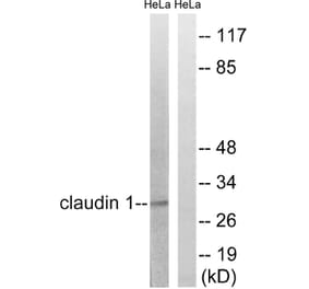
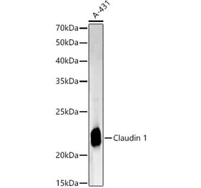
![Western Blot - Anti-Claudin 1 Antibody [ARC54478] (A81217) - Antibodies.com](https://cdn.antibodies.com/image/catalog/81/A81217_1.jpg?profile=product_alternative)