Synthetic peptide derived from human Cyclin E1 (amino acids 91-140).
Hôte
Rabbit
Clonalité
Polyclonal
Isotype
IgG
Conjuguer
Unconjugated
Purification
Purified from rabbit serum by antigen affinity chromatography using the immunizing peptide.
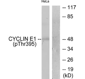
Masse moléculaire
47kDa
Forme du produit
Liquid
Formulation
Supplied in Phosphate Buffered Saline (without Mg2+ and Ca2+), pH 7.4, with 150mM NaCl, 0.02% Sodium Azide, and 50% Glycerol.
Stockage
Shipped at 4°C. Upon delivery aliquot and store at -20°C. Avoid freeze / thaw cycles.
Synonymes
CCNE, Ccne1, CCNE1_HUMAN, cyclin E variant ex5del, cyclin E variant ex7del, Cyclin Es, Cyclin Et, CyclinE, G1/S specific cyclin E, G1/S-specific cyclin-E1
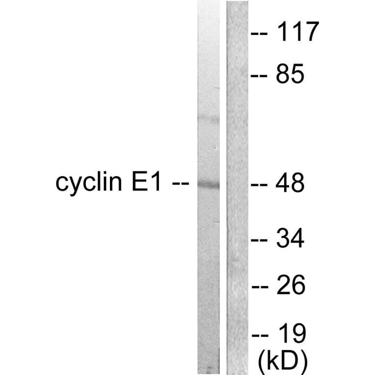
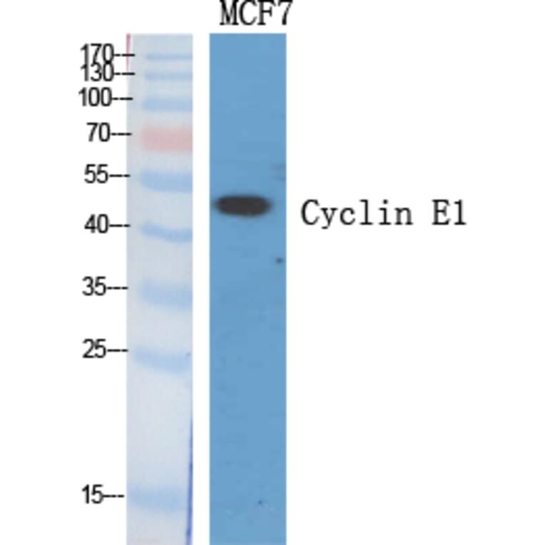
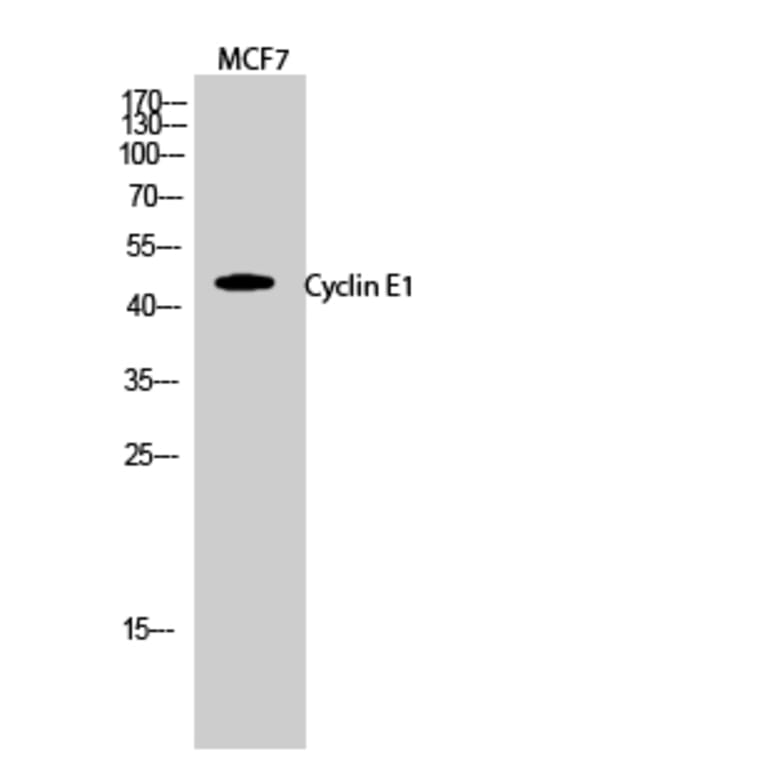
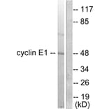
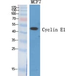
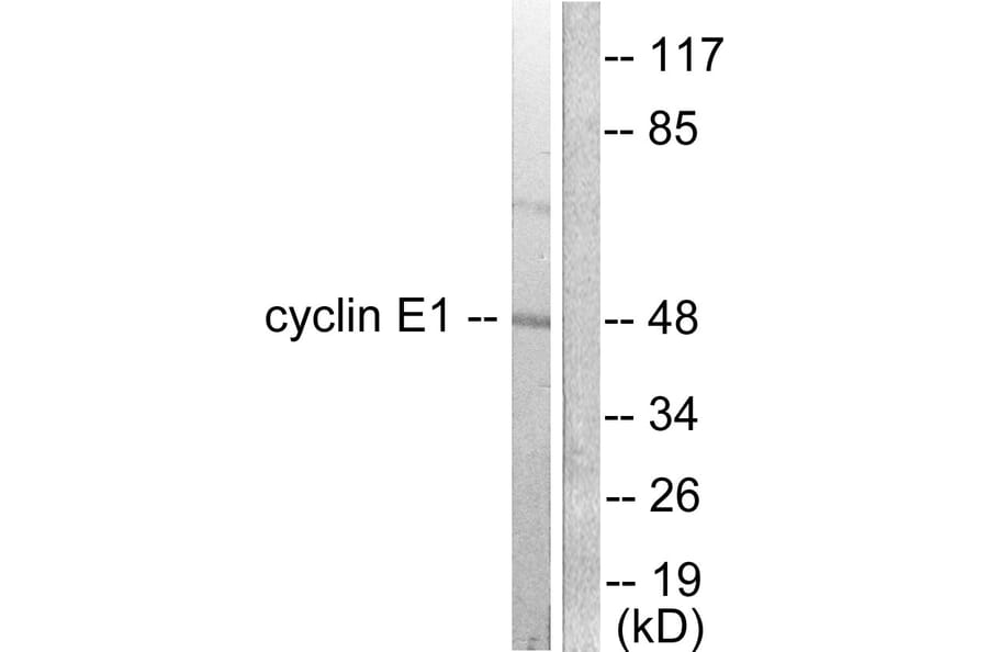
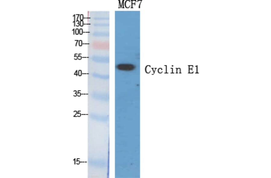
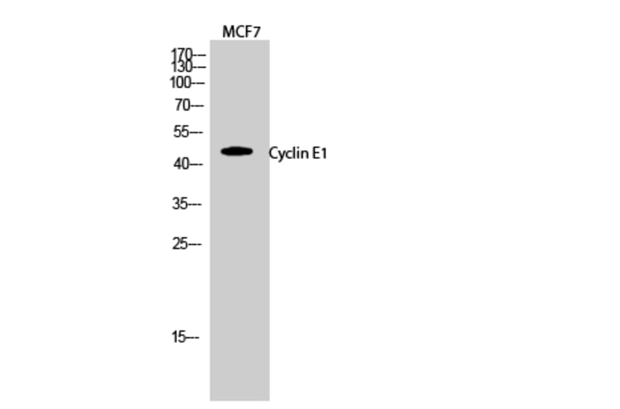
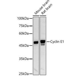
Figure 1: Western Blot - Anti-Cyclin E1 Antibody (A94771)
Western blot analysis of lysates from K562 cells using Anti-Cyclin E1 Antibody. The right hand lane represents a negative control, where the antibody is blocked by the immunising peptide.
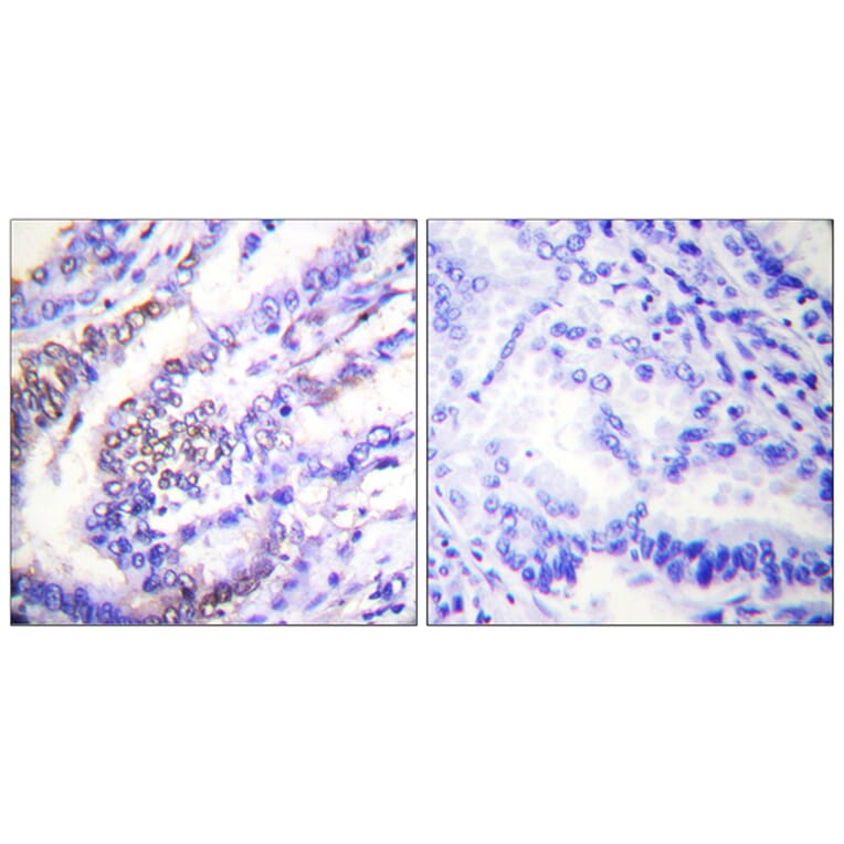
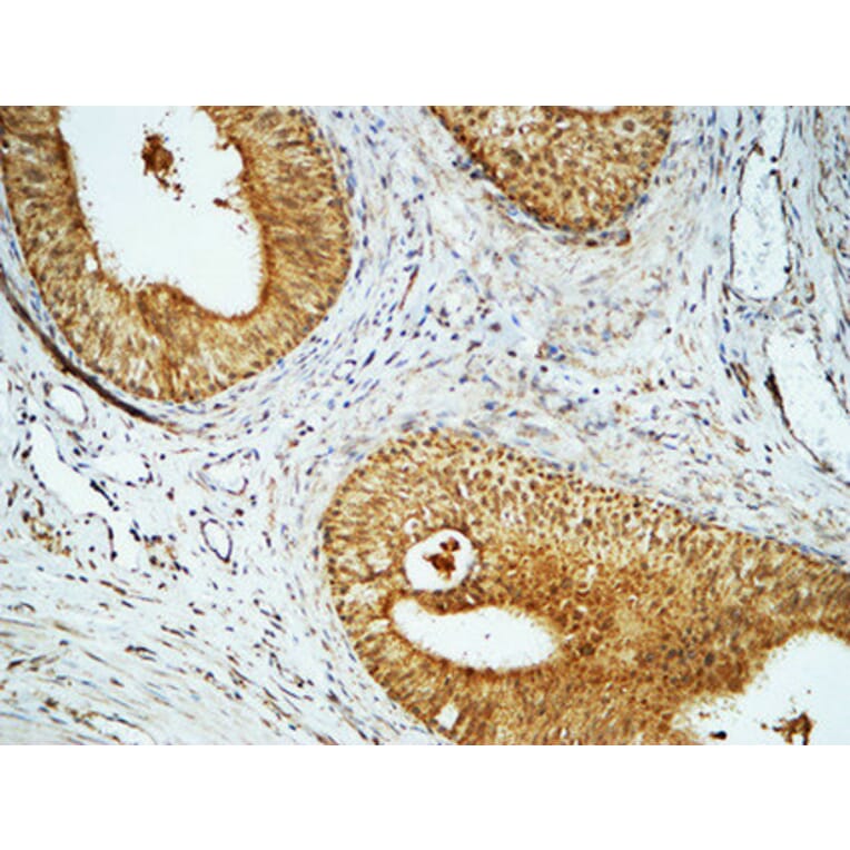
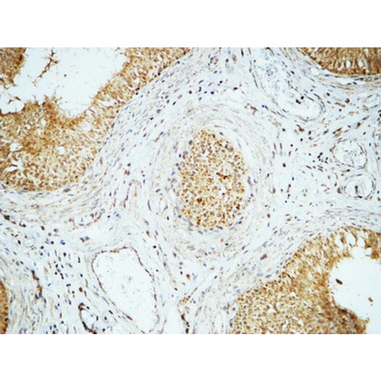
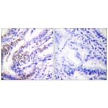
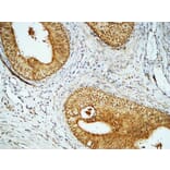
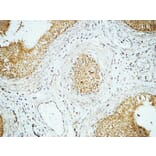
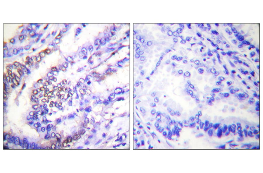
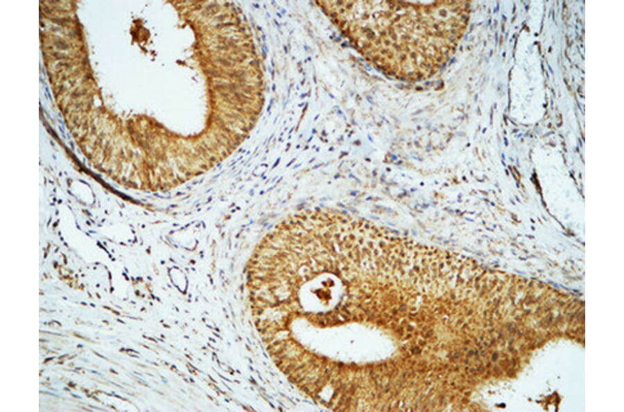
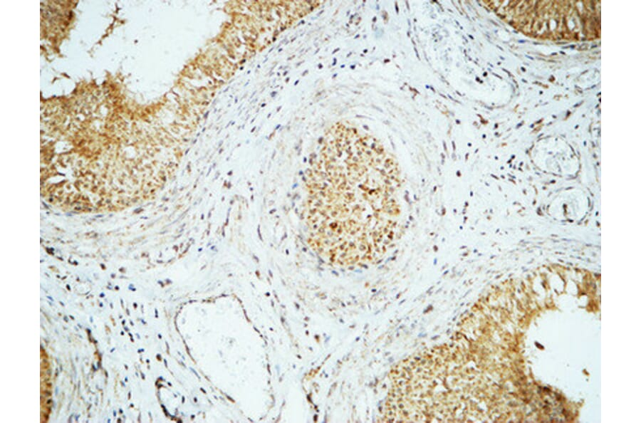
Immunohistochemical analysis of paraffin-embedded human lung carcinoma tissue using Anti-Cyclin E1 Antibody. The right hand panel represents a negative control, where the antibody was pre-incubated with the immunising peptide.
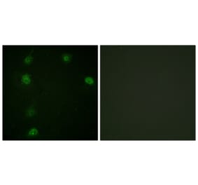
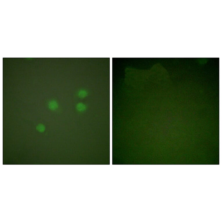
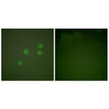
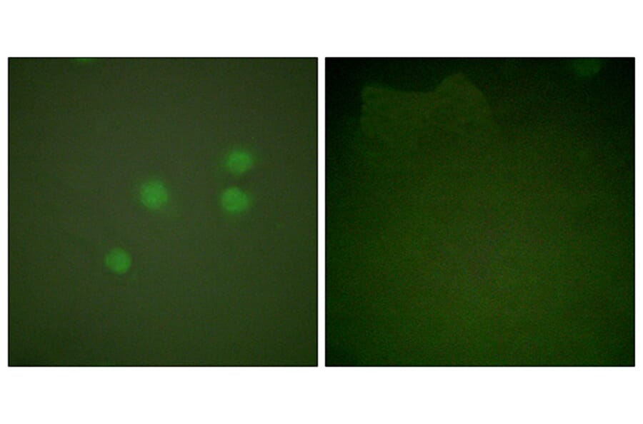
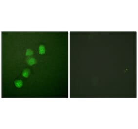
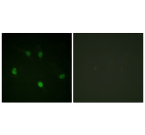
Immunofluorescence analysis of A549 cells using Anti-Cyclin E1 Antibody. The right hand panel represents a negative control, where the antibody was pre-incubated with the immunising peptide.
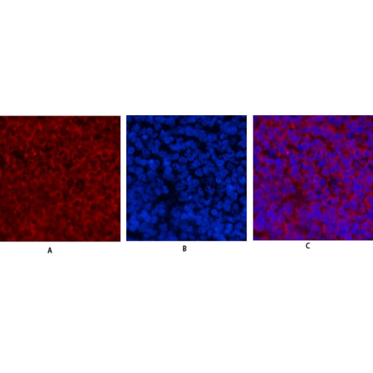
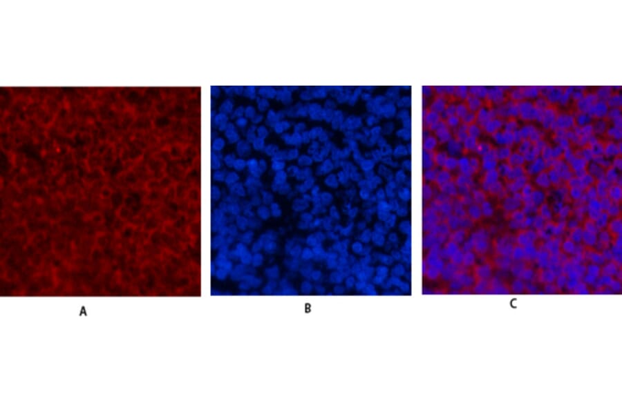
Immunofluorescence analysis of rat spleen tissue using Anti-Cyclin E1 Antibody (red) at 1:200 (4°C overnight). Cy3 labelled secondary antibody was used at 1:300 (RT 50min). Panel A: Target. Panel B: DAPI. Panel C: Merge.
Immunofluorescence analysis of rat spleen tissue using Anti-Cyclin E1 Antibody (red) at 1:200 (4°C overnight). Cy3 labelled secondary antibody was used at 1:300 (RT 50min). Panel A: Target. Panel B: DAPI. Panel C: Merge.


![Western Blot - Anti-Cyclin E1 Antibody [ARC51383] (A305272) - Antibodies.com](https://cdn.antibodies.com/image/catalog/305/A305272_1.jpg?profile=product_alternative)
![Immunohistochemistry - Anti-Cyclin E1 Antibody [rCCNE1/4936] (A277931) - Antibodies.com](https://cdn.antibodies.com/image/catalog/277/A277931_1.jpg?profile=product_alternative)
![Immunohistochemistry - Anti-Cyclin E1 Antibody [rCCNE1/4936] - BSA and Azide free (A278519) - Antibodies.com](https://cdn.antibodies.com/image/catalog/278/A278519_1.jpg?profile=product_alternative)

![Western Blot - Anti-Cyclin E1 Antibody [ARC51384 + ARC51385] (A305274) - Antibodies.com](https://cdn.antibodies.com/image/catalog/305/A305274_1.jpg?profile=product_alternative)
![Western Blot - Anti-Cyclin E1 (phospho Thr77) Antibody [ARC1559] (A306326) - Antibodies.com](https://cdn.antibodies.com/image/catalog/306/A306326_1.jpg?profile=product_alternative)