

Unconjugated
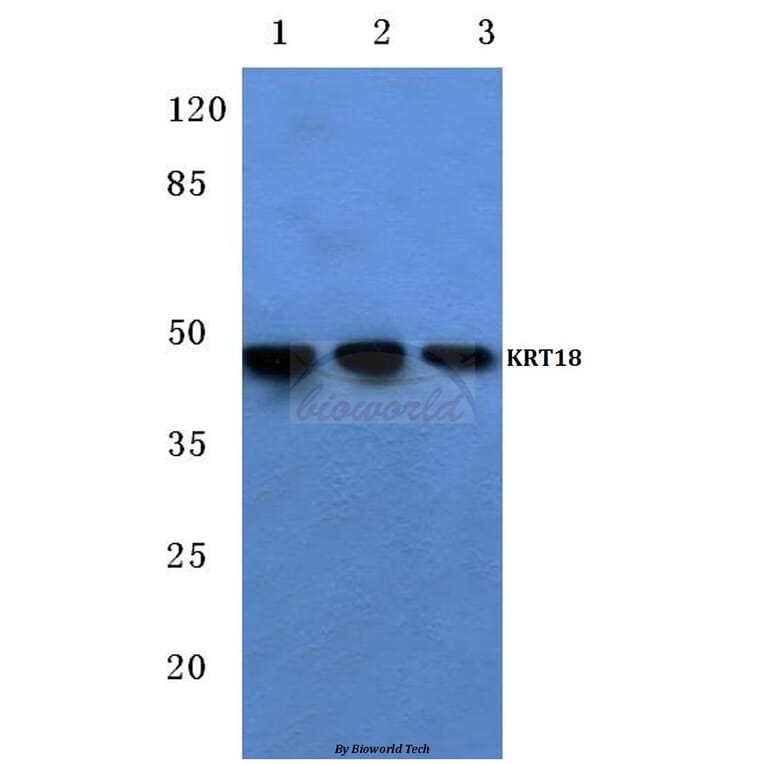
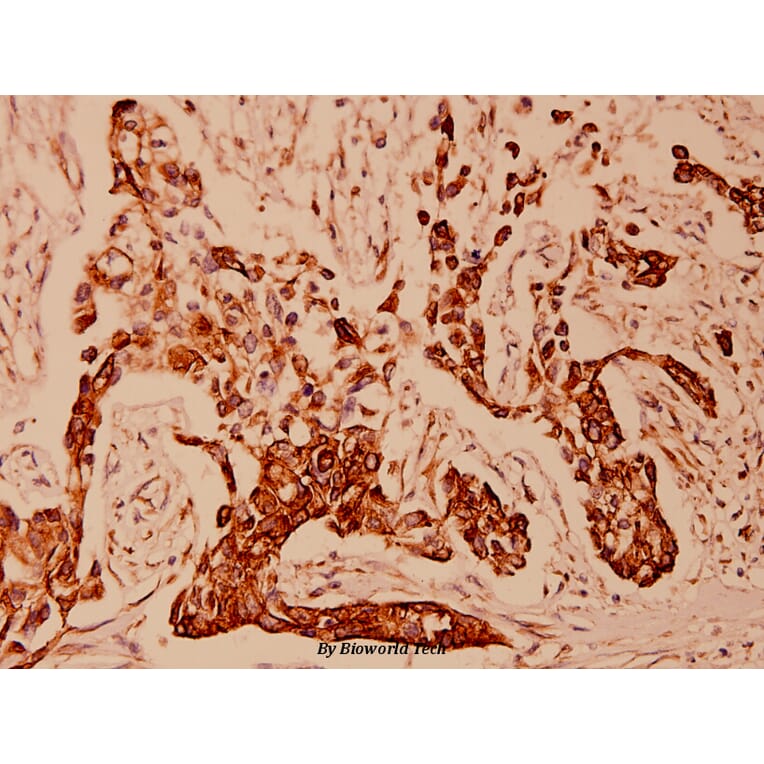
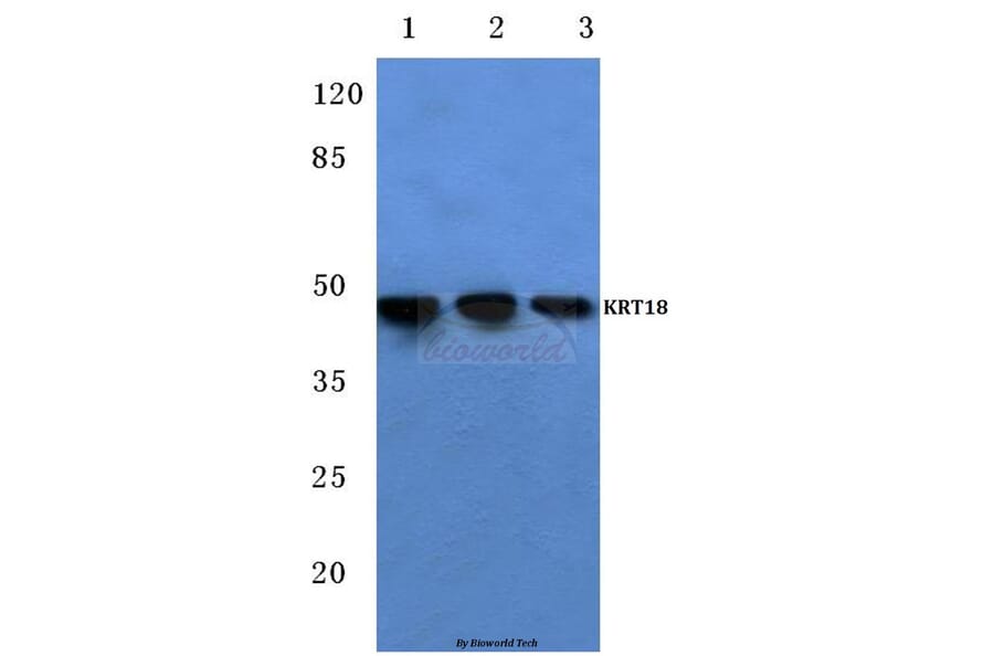
Epidermal growth factor receptor (EGFR) overexpression and activation result in increased proliferation and migration of solid tumors including ovarian cancer. In recent years, mounting evidence indicates that EGFR is a direct and functional target of miR-7. In this study, we found that miR-7 expression was significantly downregulated in highly metastatic epithelial ovarian cancer (EOC) cell lines and metastatic tissues, whereas the expression of, EGFR correlated positively with metastasis in both EOC patients and cell lines. Overexpression of miR-7 markedly suppressed the capacities of cell invasion and migration and resulted in morphological changes from a mesenchymal phenotype to an epithelial-like phenotype in EOC. In addition, overexpression of miR-7 upregulated CK-18 and β-catenin expression and downregulated Vimentin expression, accompanied with EGFR inhibition and AKT/ERK1/2 inactivation. Similar to miR-7 transfection, silencing of EGFR with this siRNA in EOC cells also upregulated CK-18 and β-catenin expression and downregulated Vimentin expression, and decreased phosphorylation of both Akt and ERK1/2, confirming that EGFR is a target of miR-7 in reversing EMT. The pharmacological inhibition of PI3K-AKT and ERK1/2 both significantly enhanced CK-18 and β-catenin expression and suppressed vimentin expression, indicating that AKT and ERK1/2 pathways are required for miR-7 mediating EMT. Finally, the expression of miR-7 and EGFR in primary EOC with matched metastasis tissues was explored. It was showed that miR-7 is inversely correlated with EGFR. Taken together, our results suggested that miR-7 inhibited tumor metastasis and reversed EMT through AKT and ERK1/2 pathway inactivation by reducing EGFR expression in EOC cell lines. Thus, miR-7 might be a potential prognostic marker and therapeutic target for ovarian cancer metastasis intervention.
Microvillous cells of the main olfactory epithelium have been described variously as primary olfactory neurons, secondary chemosensory cells or non-sensory cells. Here we generated an IP3R3(tm1(tauGFP)) mouse in which the coding region for a fusion protein of tau and green fluorescent protein replaces the first exon of the Itpr3 gene. We provide immunohistochemical and functional characterization of the cells expressing IP3 receptor type 3 in the olfactory epithelium. These cells bear microvilli at their apex, and we therefore termed them IP3R3 MV cells. The cell body of these IP3R3 MV cells lies in the upper third of the main olfactory epithelium; a long thick basal process projects towards the base of the epithelium without penetrating the basal lamina. Retrograde labeling and unilateral bulbectomy corroborated that these IP3R3 MV cells do not extend axons to the olfactory bulb and therefore are not olfactory sensory neurons. The immunohistochemical features of IP3R3 MV cells varied, suggesting either developmental stages or the existence of subsets of these cells. Thus, for example, subsets of the IP3R3 MV cells make contact with substance P fibers or express the purinergic receptor P2X3. In addition, in recordings of intracellular calcium, these cells respond to ATP and substance P as well as to a variety of odors. The characterization of IP3R3 MV cells as non-neuronal chemoresponsive cells helps to explain the differing descriptions of microvillous cells in the literature.


![Immunohistochemistry - Anti-Cytokeratin 18 Antibody [C-04] (A86656) - Antibodies.com](https://cdn.antibodies.com/image/catalog/86/A86656_794.jpg?profile=product_alternative)
![Immunohistochemistry - Anti-Cytokeratin 18 Antibody [C-04] (A249202) - Antibodies.com](https://cdn.antibodies.com/image/catalog/249/A249202_1.jpg?profile=product_alternative)
![Immunohistochemistry - Anti-Cytokeratin 18 Antibody [C-04] - BSA and Azide free (A252382) - Antibodies.com](https://cdn.antibodies.com/image/catalog/252/A252382_1.jpg?profile=product_alternative)
![Immunohistochemistry - Anti-Cytokeratin 18 Antibody [KRT18/2819R] (A249206) - Antibodies.com](https://cdn.antibodies.com/image/catalog/249/A249206_1.jpg?profile=product_alternative)
![Immunohistochemistry - Anti-Cytokeratin 18 Antibody [KRT18/2819R] - BSA and Azide free (A252386) - Antibodies.com](https://cdn.antibodies.com/image/catalog/252/A252386_1.jpg?profile=product_alternative)
![Immunohistochemistry - Anti-Cytokeratin 18 Antibody [rKRT18/1190] (A249193) - Antibodies.com](https://cdn.antibodies.com/image/catalog/249/A249193_1.jpg?profile=product_alternative)
![Immunohistochemistry - Anti-Cytokeratin 18 Antibody [rKRT18/1190] - BSA and Azide free (A252373) - Antibodies.com](https://cdn.antibodies.com/image/catalog/252/A252373_1.jpg?profile=product_alternative)
![Immunohistochemistry - Anti-Cytokeratin 18 Antibody [KRT18/834] (A249199) - Antibodies.com](https://cdn.antibodies.com/image/catalog/249/A249199_1.jpg?profile=product_alternative)
![Immunohistochemistry - Anti-Cytokeratin 18 Antibody [KRT18/834] - BSA and Azide free (A252379) - Antibodies.com](https://cdn.antibodies.com/image/catalog/252/A252379_1.jpg?profile=product_alternative)
![Immunohistochemistry - Anti-Cytokeratin 18 Antibody [DC-10] (A86578) - Antibodies.com](https://cdn.antibodies.com/image/catalog/86/A86578_740.jpg?profile=product_alternative)
![Immunohistochemistry - Anti-Cytokeratin 18 Antibody [DE-K18] - BSA and Azide free (A252378) - Antibodies.com](https://cdn.antibodies.com/image/catalog/252/A252378_1.jpg?profile=product_alternative)
