Unconjugated
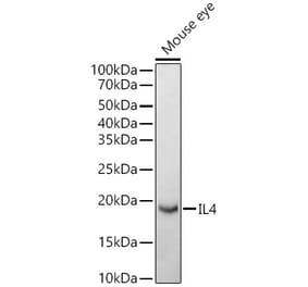
The thermally dimorphic fungus Paracoccidioides brasiliensis (Pb) is the causative agent of paracoccidioidomycosis (PCM), one of the most frequent systemic mycosis that affects the rural population in Latin America. PCM is characterized by a chronic inflammatory granulomatous reaction, which is consequence of a Th1-mediated adaptive immune response. In the present study we investigated the mechanisms involved in the immunoregulation triggered after a prior contact with cell-free antigens (CFA) during a murine model of PCM. The results showed that the inoculation of CFA prior to the infection resulted in disorganized granulomatous lesions and increased fungal replication in the lungs, liver and spleen, that paralleled with the higher levels of IL-4 when compared with the control group. The role of IL-4 in facilitating the fungal growth was demonstrated in IL-4-deficient- and neutralizing anti-IL-4 mAb-treated mice. The injection of CFA did not affect the fungal growth in these mice, which, in fact, exhibited a significant diminished amount of fungus in the tissues and smaller granulomas. Considering that in vivo anti-IL-4-application started one week after the CFA-inoculum, it implicates that IL-4-CFA-induced is responsible by the mediation of the observed unresponsiveness. Further, the characterization of CFA indicated that a proteic fraction is required for triggering the immunosuppressive mechanisms, while glycosylation or glycosphingolipids moieties are not. Taken together, our data suggest that the prior contact with soluble Pb antigens leads to severe PCM in an IL-4 dependent manner.
CD1d-restricted natural killer T (NKT) cells can rapidly produce T helper type 1 (Th1) and Th2 cytokines and also play regulatory or pathological roles in immune responses. NKT cells are able to expand when cultured with alpha-galactosylceramide (alpha-GalCer) and interleukin (IL)-2 in a CD1d-restricted manner. However, the expansion ratio of human NKT cells is variable from sample to sample. In this study, we sought to determine what factor or factors are responsible for efficient in vitro expansion of NKT cells from various inbred mouse strains. Although the proportion of NKT cells in the spleen was nearly identical in each mouse strain, the growth rates of NKT cells cultured in vitro with alpha-GalCer and IL-2 were highly variable. NKT cells from the B6C3F1 and BDF1 mouse strains expanded more than 20-fold after 4 days in culture. In contrast, NKT cells from the strain C3H/HeN did not proliferate at all. We found that cell expansion efficiency correlated with the level of IL-4 detectable in the supernatant after culture. Furthermore, we found that exogenous IL-4 augmented NKT cell proliferation early in the culture period, whereas interferon (IFN)-gamma tended to inhibit NKT cell proliferation. Thus, the ratio of production of IL-4 and IFN-gamma was important for NKT cell expansion but the absolute levels of these cytokines did not affect expansion. This finding suggests that effective expansion of NKT cells requires Th2-biased culture conditions.


![SDS-PAGE - Anti-IL-4 Antibody [11B11] (A251035) - Antibodies.com](https://cdn.antibodies.com/image/catalog/251/A251037_1.jpg?profile=product_alternative)
![SDS-PAGE - Anti-IL-4 Antibody [11B11] - BSA and Azide free (A254214) - Antibodies.com](https://cdn.antibodies.com/image/catalog/254/A254216_1.jpg?profile=product_alternative)