Synapses are specialised structures created at neuronal junctions to facilitate the transmission of electrical impulses within the nervous system. Encompassing the axon terminus of the pre-synaptic neuron, the dendrites of the post-synaptic neuron, and the synaptic cleft across which neurotransmitters diffuse, synapses connect intricate neuronal networks necessary for accurate and timely. As action potentials propagate towards the pre-synaptic axon terminus, depolarisation of the neuronal membrane triggers the opening of voltage-gated calcium channels. Subsequent Ca2+ influx signals the delivery and fusion of synaptic vesicles with the pre-synaptic membrane for neurotransmitter release. Synaptophysin and synapsin-1 are protein markers commonly used to study pre-synaptic terminals. Synaptophysin, a transmembrane protein, is involved in regulating the release of neurotransmitters while synapsin-1 helps control the reserve pool of synaptic vesicles. Several other makers involved in regulating these steps include VAMP2, the calcium-binding protein synaptotagmin, syntaxin-1, SNAP-25, and complexin-1/2. Syntaxin-1 and SNAP-25 are SNARE proteins involved in vesicle docking and fusion while complexin-1/2 interacts with SNARE complexes again to modulate neurotransmitter release. Once released into the synaptic cleft neurotransmitters diffuse towards the dendritic spines of the post-synaptic neuron. Here they are detected by receptors found grouped together within specialised zones termed the post-synaptic density (PSD). The PSD lies in close apposition to the pre-synaptic ‘active zone’, the site of neurotransmitter release. This minimises the distance across which neurotransmitters must diffuse, so increasing the speed of synaptic transmission. The marker protein bassoon is a large scaffolding protein found in the pre-synaptic active zone. PSD95, neuroligin-1 and the SHANK family of proteins all localise to the post-synaptic compartment. PSD95 is a scaffolding protein organises and stabilises postsynaptic receptors whereas neuroligin-1 is a cell adhesion molecule necessary for synapse formation. Finally, SHANK1-3 scaffolding proteins interact with postsynaptic receptors and other signalling proteins to modulate synapse development. The binding of neurotransmitters to their receptors leads to the opening or closing of ion channels in the post-synaptic neuron. Depending on the nature of the transmitter-receptor complex, these events generate postsynaptic potentials which may be excitatory or inhibitory. In this way, synaptic markers are valuable tools for visualising and monitoring the presence and distinct activity of excitatory or inhibitory neurotransmitters. Transporter proteins VGLUT1-3 act as markers for vesicular loading of the excitatory neurotransmitter glutamate. Similarly, the vesicular GABA transporter (VGAT) loads GABA and glycine into synaptic vesicles from the neuronal cytoplasm. GABA is the primary inhibitory neurotransmitter in the brain and can also be detected directly to identify inhibitory synapses. Other markers such as synGAP and the adhesion protein GAP43 play vital roles regulating the strength and plasticity of neuronal connections. Understanding the functions and interactions of these markers is crucial for unravelling the mechanistic basis of communication. We offer a range of antibodies against synaptic markers including Neuroligin 1 antibodies and Synapsin I antibodies, that are validated across multiple applications and cover various host species, isotypes and conjugates.
Anti-Synaptophysin Antibody (A54004) | |
100µg/$595 | |
| Description: | Rabbit polyclonal antibody to Synaptophysin |
| Applications: | CM, ELISA, ICC, IF, IHC, IP, WB |
| Reactivity: | Chicken, Human, Monkey, Mouse, Rat |
| Conjugate: | Unconjugated |
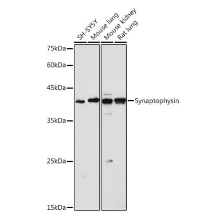
Anti-Synaptophysin Antibody (A15183) | |
100µl/$375 | |
| Description: | Rabbit polyclonal antibody to Synaptophysin. |
| Applications: | WB, IHC, ICC/IF, IP |
| Reactivity: | Human, Mouse, Rat |
| Conjugate: | Unconjugated |
Anti-PSD95 Antibody [rK28/43] (CF®647) (A352071) | |
500µl/$660 | |
| Description: | Recombinant mouse monoclonal [rK28/43] antibody to PSD95 (CF®647). |
| Applications: | IF, IHC, IHC-P, WB |
| Reactivity: | Human, Mouse, Rat, Chicken, Non-Human Primates |
| Conjugate: | CF®647 |
![Immunofluorescence - Anti-PSD95 Antibody [rK28/43] (CF®647) (A352071) - Antibodies.com](https://cdn.antibodies.com/image/catalog/352/A352071_1.webp?profile=product_search)
Anti-PSD95 Antibody [rK28/43] (CF®680R) (A352072) | |
500µl/$685 | |
| Description: | Recombinant mouse monoclonal [rK28/43] antibody to PSD95 (CF®680R). |
| Applications: | IF, IHC, IHC-P, WB |
| Reactivity: | Human, Mouse, Rat, Chicken, Non-Human Primates |
| Conjugate: | CF®680R |
![Immunofluorescence - Anti-PSD95 Antibody [rK28/43] (CF®680R) (A352072) - Antibodies.com](https://cdn.antibodies.com/image/catalog/352/A352072_1.webp?profile=product_search)
Anti-PSD95 Antibody [rK28/43] (CF®740) (A352073) | |
500µl/$685 | |
| Description: | Recombinant mouse monoclonal [rK28/43] antibody to PSD95 (CF®740). |
| Applications: | IF, IHC, IHC-P, WB |
| Reactivity: | Human, Mouse, Rat, Chicken, Non-Human Primates |
| Conjugate: | CF®740 |
Anti-PSD95 Antibody [rK28/43] (CF®790) (A352074) | |
500µl/$720 | |
| Description: | Recombinant mouse monoclonal [rK28/43] antibody to PSD95 (CF®790). |
| Applications: | IF, IHC, IHC-P, WB |
| Reactivity: | Human, Mouse, Rat, Chicken, Non-Human Primates |
| Conjugate: | CF®790 |
Anti-Synapsin Antibody (A96306) | |
10µg - 100µg/$200 – $505 | |
| Description: | Rabbit polyclonal antibody to Synapsin. |
| Applications: | WB, IHC, IF, ELISA |
| Reactivity: | Human, Mouse, Rat |
| Conjugate: | Unconjugated |
Anti-PSD95 Antibody [rK28/43] (CF®488A) (A352067) | |
500µl/$660 | |
| Description: | Recombinant mouse monoclonal [rK28/43] antibody to PSD95 (CF®488A). |
| Applications: | IF, IHC, IHC-P, WB |
| Reactivity: | Human, Mouse, Rat, Chicken, Non-Human Primates |
| Conjugate: | CF®488A |
Anti-PSD95 Antibody [rK28/43] (CF®568) (A352068) | |
500µl/$660 | |
| Description: | Recombinant mouse monoclonal [rK28/43] antibody to PSD95 (CF®568). |
| Applications: | IF, IHC, IHC-P, WB |
| Reactivity: | Human, Mouse, Rat, Chicken, Non-Human Primates |
| Conjugate: | CF®568 |
Anti-PSD95 Antibody [rK28/43] (CF®594) (A352069) | |
500µl/$660 | |
| Description: | Recombinant mouse monoclonal [rK28/43] antibody to PSD95 (CF®594). |
| Applications: | IF, IHC, IHC-P, WB |
| Reactivity: | Human, Mouse, Rat, Chicken, Non-Human Primates |
| Conjugate: | CF®594 |
Anti-PSD95 Antibody [rK28/43] (CF®640R) (A352070) | |
500µl/$660 | |
| Description: | Recombinant mouse monoclonal [rK28/43] antibody to PSD95 (CF®640R). |
| Applications: | IF, IHC, IHC-P, WB |
| Reactivity: | Human, Mouse, Rat, Chicken, Non-Human Primates |
| Conjugate: | CF®640R |
Anti-Synaptophysin Antibody (FITC) (A52142) | |
100µg/$655 | |
| Description: | Rabbit polyclonal antibody to Synaptophysin (FITC) |
| Applications: | CM, ELISA, ICC, IF, IHC, IP, WB |
| Reactivity: | Chicken, Human, Monkey, Mouse, Rat |
| Conjugate: | FITC |
Anti-Synaptophysin Antibody (Biotin) (A54355) | |
100µg/$655 | |
| Description: | Rabbit polyclonal antibody to Synaptophysin (Biotin) |
| Applications: | CM, ELISA, ICC, IF, IHC, IMM, WB |
| Reactivity: | Chicken, Human, Monkey, Mouse, Rat |
| Conjugate: | Biotin |
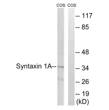
Anti-Syntaxin 1A Antibody (A94334) | |
10µg - 100µg/$200 – $505 | |
| Description: | Rabbit polyclonal antibody to Syntaxin 1A. |
| Applications: | WB, IHC, IF, ELISA |
| Reactivity: | Human, Mouse, Rat, Monkey |
| Conjugate: | Unconjugated |
Anti-PSD95 Antibody [7E3] (A304702) | |
100µg/$510 | |
| Description: | Mouse monoclonal [7E3] antibody to PSD95. |
| Applications: | WB, IHC, ICC/IF, Antibody Microarray |
| Reactivity: | Human, Mouse, Rat, Bovine |
| Conjugate: | Unconjugated |
Anti-Synaptophysin Antibody (FITC) (A52143) | |
100µg/$655 | |
| Description: | Rabbit polyclonal antibody to Synaptophysin (FITC) |
| Applications: | CM, ELISA, ICC, IF, IHC, IMM, WB |
| Reactivity: | Chicken, Human, Monkey, Mouse, Rat |
| Conjugate: | FITC |
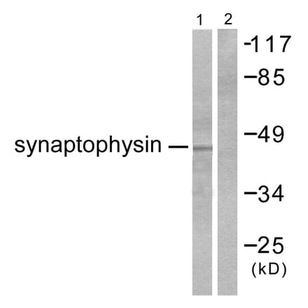
Anti-Synaptophysin Antibody (A54005) | |
100µg/$595 | |
| Description: | Rabbit polyclonal antibody to Synaptophysin |
| Applications: | CM, ELISA, ICC, IF, IHC, IMM, WB |
| Reactivity: | Chicken, Human, Monkey, Mouse, Rat |
| Conjugate: | Unconjugated |
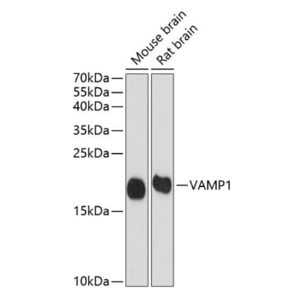
Anti-VAMP1 Antibody (A16275) | |
100µl/$375 | |
| Description: | Rabbit polyclonal antibody to VAMP1. |
| Applications: | WB, IHC, ICC/IF |
| Reactivity: | Human, Mouse, Rat |
| Conjugate: | Unconjugated |
Anti-Synaptophysin Antibody (Biotin) (A54354) | |
100µg/$655 | |
| Description: | Rabbit polyclonal antibody to Synaptophysin (Biotin) |
| Applications: | CM, ELISA, ICC, IF, IHC, IP, WB |
| Reactivity: | Chicken, Human, Monkey, Mouse, Rat |
| Conjugate: | Biotin |
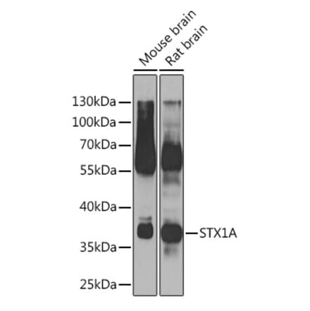
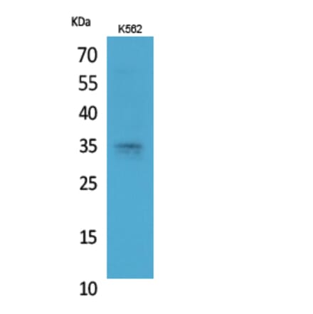
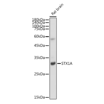
Anti-STX1A Antibody (A97182) | |
10µg - 100µg/$200 – $505 | |
| Description: | Rabbit polyclonal antibody to STX1A. |
| Applications: | WB, IHC, IF, ELISA |
| Reactivity: | Human, Mouse, Rat |
| Conjugate: | Unconjugated |
Anti-PSD95 Antibody [6G6] (A304703) | |
100µg/$510 | |
| Description: | Mouse monoclonal [6G6] antibody to PSD95. |
| Applications: | WB, IHC, ICC/IF, Antibody Microarray |
| Reactivity: | Human, Mouse, Rat, Bovine |
| Conjugate: | Unconjugated |
Anti-Syntaxin 1a Antibody [ARC2403] (A307451) | |
100µl/$515 | |
| Description: | Rabbit monoclonal [ARC2403] antibody to Syntaxin 1a. |
| Applications: | WB, IHC, ICC/IF |
| Reactivity: | Human, Mouse, Rat |
| Conjugate: | Unconjugated |
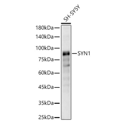
Anti-Synapsin1 Antibody (A95172) | |
10µg - 100µg/$200 – $505 | |
| Description: | Rabbit polyclonal antibody to Synapsin1. |
| Applications: | WB, IHC, IF, ELISA |
| Reactivity: | Human, Mouse, Rat |
| Conjugate: | Unconjugated |
Anti-PSD95 Antibody (A82926) | |
100µg/$525 | |
| Description: | Goat polyclonal antibody to PSD95. |
| Applications: | ELISA, WB |
| Reactivity: | Rat |
| Conjugate: | Unconjugated |
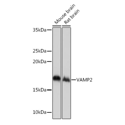
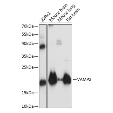
Anti-VAMP2 Antibody (A13407) | |
100µl/$375 | |
| Description: | Rabbit polyclonal antibody to VAMP2. |
| Applications: | WB, IHC |
| Reactivity: | Human, Mouse, Rat |
| Conjugate: | Unconjugated |
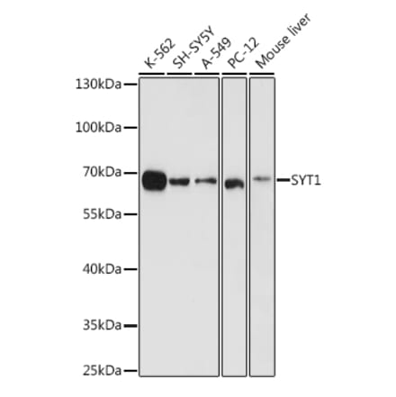
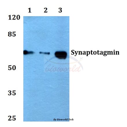
Anti-Synaptotagmin Antibody (A15863) | |
100µl/$375 | |
| Description: | Rabbit polyclonal antibody to Synaptotagmin. |
| Applications: | WB, IHC, ICC/IF |
| Reactivity: | Human, Mouse, Rat |
| Conjugate: | Unconjugated |
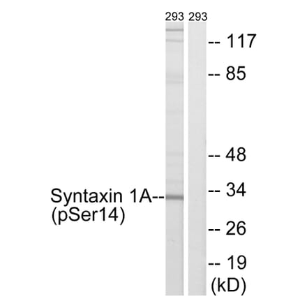
Anti-Syntaxin 1A (phospho Ser14) Antibody (A93452) | |
10µg - 100µg/$200 – $505 | |
| Description: | Rabbit polyclonal antibody to Syntaxin 1A (phospho Ser14). |
| Applications: | WB, IHC, IF, ELISA |
| Reactivity: | Human, Mouse, Rat |
| Conjugate: | Unconjugated |
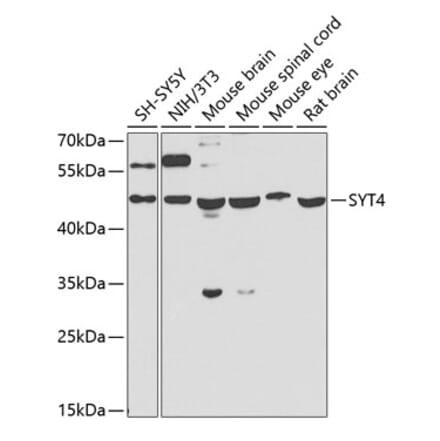
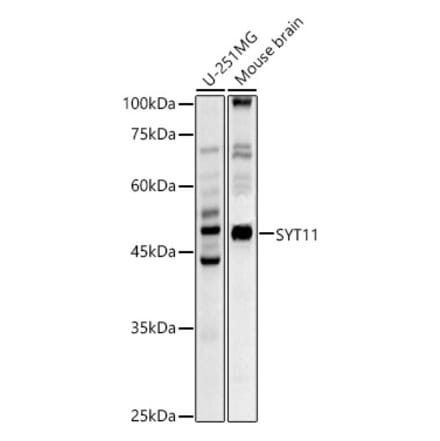
Anti-SYT11 Antibody (A16111) | |
100µl/$375 | |
| Description: | Rabbit polyclonal antibody to SYT11. |
| Applications: | WB, ICC/IF |
| Reactivity: | Human, Mouse, Rat |
| Conjugate: | Unconjugated |
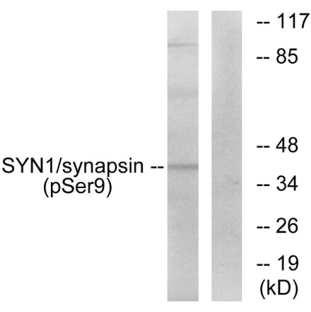
Anti-Synapsin (phospho Ser9) Antibody (A93658) | |
10µg - 100µg/$200 – $505 | |
| Description: | Rabbit polyclonal antibody to Synapsin (phospho Ser9). |
| Applications: | WB, IHC, IF, ELISA |
| Reactivity: | Human, Mouse, Rat |
| Conjugate: | Unconjugated |
Anti-Synaptophysin Antibody (A94747) | |
10µg - 100µg/$200 – $505 | |
| Description: | Rabbit polyclonal antibody to Synaptophysin. |
| Applications: | WB, IHC, IF, ELISA |
| Reactivity: | Human, Mouse, Rat |
| Conjugate: | Unconjugated |
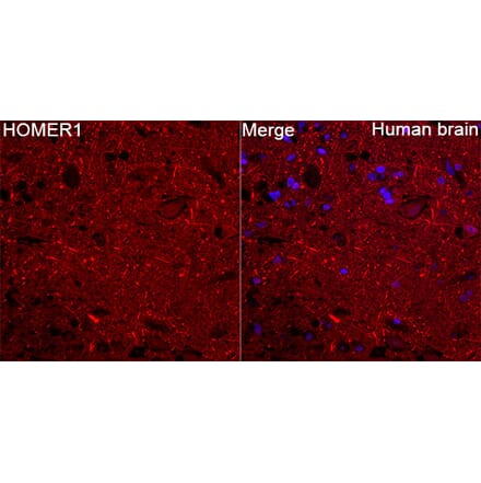
Anti-HOMER1 Antibody (A9430) | |
100µl/$375 | |
| Description: | Rabbit polyclonal antibody to HOMER1. |
| Applications: | WB, ICC/IF, ELISA |
| Reactivity: | Human, Mouse, Rat |
| Conjugate: | Unconjugated |
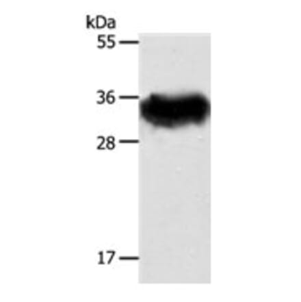
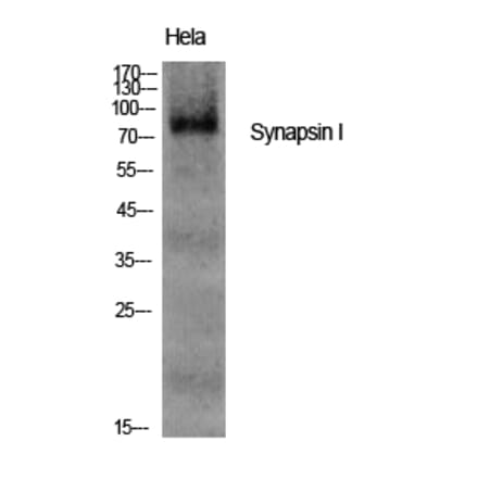
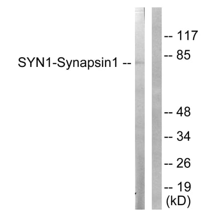
Anti-Synapsin I Antibody (A11819) | |
100µl/$375 | |
| Description: | Rabbit polyclonal antibody to Synapsin I. |
| Applications: | WB, ICC/IF |
| Reactivity: | Human, Mouse, Rat |
| Conjugate: | Unconjugated |
Anti-Neuroligin 1 Antibody [S97A-31] (A304803) | |
100µg/$550 | |
| Description: | Mouse monoclonal [S97A-31] antibody to Neuroligin 1. |
| Applications: | WB, IHC, ICC/IF |
| Reactivity: | Human, Mouse, Rat |
| Conjugate: | Unconjugated |
Anti-Synaptotagmin VII Antibody [S275-14] (A304818) | |
100µg/$550 | |
| Description: | Mouse monoclonal [S275-14] antibody to Synaptotagmin VII. |
| Applications: | WB, IHC, ICC/IF |
| Reactivity: | Human, Mouse, Rat |
| Conjugate: | Unconjugated |
Anti-Syntaxin 1a Antibody (A15983) | |
100µl/$375 | |
| Description: | Rabbit polyclonal antibody to Syntaxin 1a. |
| Applications: | WB, IHC, ICC/IF |
| Reactivity: | Mouse, Rat |
| Conjugate: | Unconjugated |
Anti-Synapsin1 (phospho Ser62) Antibody (A93607) | |
10µg - 100µg/$200 – $505 | |
| Description: | Rabbit polyclonal antibody to Synapsin1 (phospho Ser62). |
| Applications: | WB, IHC, IF, ELISA |
| Reactivity: | Human, Mouse, Rat |
| Conjugate: | Unconjugated |
Anti-VAMP2 Antibody [ARC0936] (A307718) | |
100µl/$515 | |
| Description: | Rabbit monoclonal [ARC0936] antibody to VAMP2. |
| Applications: | WB, ICC/IF |
| Reactivity: | Human, Mouse, Rat |
| Conjugate: | Unconjugated |
Anti-Synaptophysin Antibody [Q21-Q] (A8238) | |
100µl - 1ml/$455 – $1,000 | |
| Description: | Rabbit monoclonal (Q21-Q) antibody to Synaptophysin. |
| Applications: | IHC-P, IHC-Fr |
| Reactivity: | Human |
| Conjugate: | Unconjugated |
Anti-STX1A Antibody (A48160) | |
100µl/$430 | |
| Description: | Rabbit polyclonal antibody to STX1A |
| Applications: | ELISA, WB, IHC |
| Reactivity: | Human, Mouse, Rat |
| Conjugate: | Unconjugated |
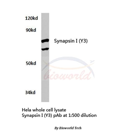
Anti-Synapsin I (Y3) Antibody (A27080) | |
50µl - 100µl/$415 – $560 | |
| Description: | Rabbit polyclonal antibody to Synapsin I (Y3) |
| Applications: | WB, IHC, IF |
| Reactivity: | Human, Mouse, Rat |
| Conjugate: | Unconjugated |
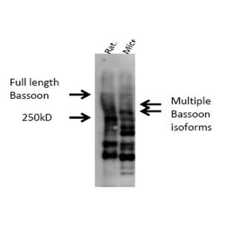
Anti-Bassoon Antibody - BSA and Azide free (A304191) | |
100µg/$745 | |
| Description: | Rabbit polyclonal antibody to Bassoon. |
| Applications: | WB, IHC, IF |
| Reactivity: | Human, Mouse, Rat |
| Conjugate: | Unconjugated |
Anti-Synaptotagmin Antibody (A12750) | |
100µl/$375 | |
| Description: | Rabbit polyclonal antibody to Synaptotagmin. |
| Applications: | WB, ICC/IF |
| Reactivity: | Human, Mouse, Rat |
| Conjugate: | Unconjugated |
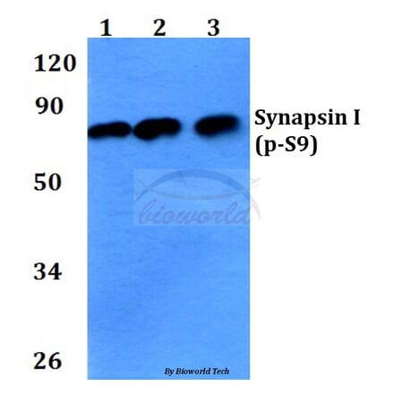
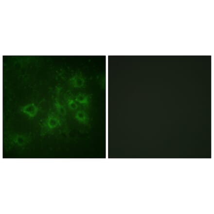
Anti-Synapsin I (phospho-S9) Antibody (A27747) | |
50µl - 100µl/$415 – $560 | |
| Description: | Rabbit polyclonal antibody to Synapsin I (phospho-S9) |
| Applications: | WB, IHC, IF |
| Reactivity: | Human, Mouse, Rat |
| Conjugate: | Unconjugated |
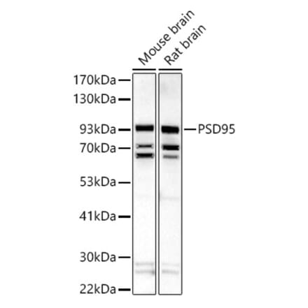
Anti-PSD95 Antibody (A15101) | |
100µl/$375 | |
| Description: | Rabbit polyclonal antibody to PSD95. |
| Applications: | WB, ICC/IF |
| Reactivity: | Human, Mouse, Rat |
| Conjugate: | Unconjugated |
Anti-Synapsin I Antibody (A34298) | |
50µl - 100µl/$290 – $430 | |
| Description: | Rabbit polyclonal antibody to Synapsin I |
| Applications: | ELISA, WB, ICC/IF, IHC |
| Reactivity: | Human, Mouse, Rat |
| Conjugate: | Unconjugated |
Anti-Synapsin1 (phospho Ser605) Antibody (A93457) | |
10µg - 100µg/$200 – $505 | |
| Description: | Rabbit polyclonal antibody to Synapsin1 (phospho Ser605). |
| Applications: | IHC, IF, ELISA |
| Reactivity: | Human, Mouse, Rat |
| Conjugate: | Unconjugated |
Anti-Synaptophysin (L128) Antibody (A25371) | |
50µl - 100µl/$415 – $560 | |
| Description: | Rabbit polyclonal antibody to Synaptophysin (L128) |
| Applications: | WB, IHC |
| Reactivity: | Human, Mouse, Rat |
| Conjugate: | Unconjugated |
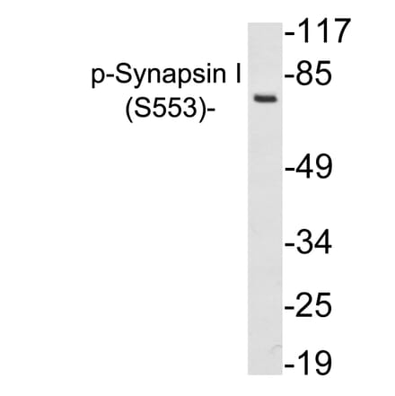
Anti-Synapsin I (phospho Ser553) Antibody (A94141) | |
10µg - 100µg/$200 – $505 | |
| Description: | Rabbit polyclonal antibody to Synapsin I (phospho Ser553). |
| Applications: | WB, IHC |
| Reactivity: | Human, Mouse, Rat |
| Conjugate: | Unconjugated |
Anti-Synaptotagmin (T196) Antibody (A25372) | |
50µl - 100µl/$415 – $560 | |
| Description: | Rabbit polyclonal antibody to Synaptotagmin (T196) |
| Applications: | WB, IHC |
| Reactivity: | Human, Mouse, Rat |
| Conjugate: | Unconjugated |
Anti-VAMP2 Antibody (A38276) | |
100µl/$430 | |
| Description: | Rabbit polyclonal antibody to VAMP2 |
| Applications: | WB, IHC |
| Reactivity: | Human, Mouse |
| Conjugate: | Unconjugated |
Showing 1-50 of 146 products
![Immunofluorescence - Anti-PSD95 Antibody [rK28/43] (CF®740) (A352073) - Antibodies.com](https://cdn.antibodies.com/image/catalog/352/A352073_1.webp?profile=product_search)
![Immunofluorescence - Anti-PSD95 Antibody [rK28/43] (CF®790) (A352074) - Antibodies.com](https://cdn.antibodies.com/image/catalog/352/A352074_1.webp?profile=product_search)

![Immunofluorescence - Anti-PSD95 Antibody [rK28/43] (CF®488A) (A352067) - Antibodies.com](https://cdn.antibodies.com/image/catalog/352/A352067_1.webp?profile=product_search)
![Immunofluorescence - Anti-PSD95 Antibody [rK28/43] (CF®568) (A352068) - Antibodies.com](https://cdn.antibodies.com/image/catalog/352/A352068_1.webp?profile=product_search)
![Immunofluorescence - Anti-PSD95 Antibody [rK28/43] (CF®594) (A352069) - Antibodies.com](https://cdn.antibodies.com/image/catalog/352/A352069_1.webp?profile=product_search)
![Immunofluorescence - Anti-PSD95 Antibody [rK28/43] (CF®640R) (A352070) - Antibodies.com](https://cdn.antibodies.com/image/catalog/352/A352070_1.webp?profile=product_search)

![Western Blot - Anti-PSD95 Antibody [7E3] (A304702) - Antibodies.com](https://cdn.antibodies.com/image/catalog/304/A304702_1.png?profile=product_search)
![Western Blot - Anti-PSD95 Antibody [6G6] (A304703) - Antibodies.com](https://cdn.antibodies.com/image/catalog/304/A304703_1.png?profile=product_search)
![Immunocytochemistry/Immunofluorescence - Anti-Neuroligin 1 Antibody [S97A-31] (A304803) - Antibodies.com](https://cdn.antibodies.com/image/catalog/304/A304803_1.png?profile=product_search)
![Immunocytochemistry/Immunofluorescence - Anti-Synaptotagmin VII Antibody [S275-14] (A304818) - Antibodies.com](https://cdn.antibodies.com/image/catalog/304/A304818_1.png?profile=product_search)