ELISA Protocols

By Ryan Hamnett, PhD

ELISAs use antibodies to detect and quantify the amount of a target antigen in a liquid sample. Below are example protocols covering all the stages of multiple ELISA formats and assaying various types of sample for the detection of target proteins.

Following processing, all sample types should be aliquoted to minimize freeze-thaw cycles, and stored at -80°C.

Cell culture supernatant

  • Centrifuge at 1500 rpm for 10 min at 4°C.

Cell extracts

  • Remove culture media and wash with ice-cold PBS.
  • Add extraction buffer, then scrape the cells and put into a chilled tube.
  • Vortex briefly and incubate on ice for 15-30 min.
  • Centrifuge at 13000 rpm for 10 minutes at 4°C (pellets insoluble content).

Tissue extract

  • Rinse tissue in ice-cold PBS to remove excess blood before homogenization.
  • Place into a round-bottomed tube and immerse in liquid nitrogen to snap freeze.
  • Homogenize tissue: for every 5 mg tissue, add 300 ul extraction buffer.
  • Incubate sample with homogenization buffer on a shaker for 2 hours at 4°C.
  • Centrifuge for 20 minutes at 13000 rpm at 4°C.

Serum

  • Collect blood in serum-separator tubes.
  • Allow blood to clot for 30 minutes at room temperature.
  • After clotting, centrifuge at 1000 x g for 15 minutes.

Plasma

  • Collect plasma in tube containing EDTA or heparin as an anticoagulant, or add 0.1 M sodium citrate to 1/10 final volume.
  • Centrifuge at 3000 x g for 15 minutes at 4°C within 30 minutes of collection.

Other biological fluids (e.g. milk, saliva, urine)

  • Collect sample and centrifuge at 10,000 g for 2 minutes at 4°C.

Antigen coating

  1. Coat microtiter plate wells with 100 μl of coating antigen in coating buffer.
    1. Concentration should be 2-20 μg/ml (20 μg/ml saturating).
    2. Pure solutions are not essential, but the target antigen should represent more than 3% of the protein content in the sample.
  2. Cover and incubate overnight at 4°C or for 30 minutes at 37°C.
  3. Remove coating solution by flicking plate over sink, and wash 3x 5 minutes with 200 μl wash buffer.
    1. Do not let the plate dry at any point during the protocol.

Blocking and standard preparation

  1. Add 150 μl blocking solution to each well. Cover and incubate for 1 hour at 37°C.
  2. Meanwhile, prepare standards as indicated in product manual or in the Reagents and Buffers section below.
  3. Wash 2x 5 minutes in 200 μl wash buffer.

Primary antibody incubation

  1. Add 100 μl primary antibody diluted in blocking buffer.
    1. Antibody concentration should be determined by dilution series, using manufacturer’s recommendation as a guide.
    2. For direct ELISA, the primary antibody will be conjugated to an enzyme (HRP or AP) or fluorophore. For indirect ELISA, it will be unlabeled.
  2. Cover and incubate for 2 hours at room temperature, or overnight at 4°C for a stronger signal.
  3. Wash 4x 5 minutes in 200 μl wash buffer.

Secondary antibody incubation (only for Indirect ELISA)

  1. Add 100 μl labeled secondary antibody diluted in blocking buffer.
  2. Cover and incubate for 1-2 hours at room temperature.
  3. Wash 4x 5 minutes in 200 μl wash buffer.

Detection and analysis

  1. Prepare the substrate solution appropriate for your enzyme.
    1. Before adding TMB into the wells, equilibrate TMB for 30 minutes at 37°C.
    2. Sodium azide is often used as a preservative for antibody solutions and buffers, but it should be avoided because it inhibits HRP.
  2. Add 100 μl substrate solution to each well.
  3. Cover and incubate at room temperature for 30 minutes.
    1. c. TMB is sensitive to light. Perform this incubation in the dark by wrapping the plate in foil.
  4. Read absorbance at the appropriate wavelength immediately, or add 50 μl stop solution, tapping to ensure it has thoroughly mixed, then read absorbance within 30 minutes.
  5. Generate a standard curve from the serial dilutions of standards, plotting concentration on the x axis and absorbance on the y axis.
    1. More information on analysis can be found in our ELISA Overview guide: Standard curve.

All sandwich ELISA plates sold by Antibodies.com come pre-coated with capture antibody, so the antibody coating steps of this protocol are not necessary to perform.

Antibody coating

  1. Coat microtiter plate wells with 100 μl of capture antibody in coating buffer.
    1. Concentration should be 1-10 μg/ml.
    2. Non-purified antibodies, such as antiserum, should be used at the upper end of this concentration range as the concentration of the antibody itself will be lower.
  2. Cover and incubate overnight at 4°C or for 30 minutes at 37°C.
  3. Remove coating solution by flicking plate over sink, and wash 3x 5 minutes with 200 μl wash buffer.
    1. Do not let the plate dry at any point during the protocol.

Blocking and standard preparation

  1. Add 150 μl blocking solution to each well. Cover and incubate for 1 hour at 37°C.
  2. Meanwhile, prepare standards as indicated in product manual or in the Reagents and Buffers section below.
  3. Wash 2x 5 minutes in 200 μl wash buffer.

Sample incubation

  1. Add 100 μl diluted sample to each well.
    1. Samples should be diluted such that the analyte concentration falls within the linear detection range of the assay.
    2. Samples should be run in duplicate or triplicate to ensure accuracy.
  2. Cover and incubate for 90 minutes at room temperature, or overnight at 4°C.
  3. Wash 4x 5 minutes in 200 μl wash buffer.

Biotinylated antibody incubation

  1. Add 100 μl biotin-labeled detection antibody diluted in blocking buffer.
  2. Cover and incubate for 2 hours at room temperature or 1 hour at 37°C.
  3. Wash 4x 5 minutes in 200 μl wash buffer.
    1. If performing an indirect sandwich ELISA, the detection antibody will be unlabeled. After incubation and washing, a labeled secondary antibody should be added, incubated for 1-2 hours, and then washed 4 times as above.

Biotinylated antibody incubation

  1. Add 100 μl enzyme-conjugated streptavidin (appropriately diluted in wash buffer) to each well.
  2. Cover and incubate for 30-60 minutes at 37°C.
  3. Wash 3x 5 minutes in 200 μl wash buffer.

Detection and analysis

  1. Prepare the substrate solution appropriate for your enzyme
    1. Before adding TMB into the wells, equilibrate TMB for 30 minutes at 37°C.
    2. Sodium azide is often used as a preservative for antibody solutions and buffers, but it should be avoided because it inhibits HRP.
  2. Add 100 μl substrate solution to each well.
  3. Cover and incubate at room temperature for 30 minutes.
    1. TMB is sensitive to light. Perform this incubation in the dark by wrapping the plate in foil.
  4. Read absorbance at the appropriate wavelength immediately, or add 50 μl stop solution, tapping to ensure it has thoroughly mixed, then read absorbance within 30 minutes.
  5. Generate a standard curve from the serial dilutions of standards, plotting concentration on the x axis and absorbance on the y axis.
    1. More information on analysis can be found in our ELISA Overview guide: Standard curve.

All competitive ELISA plates sold by Antibodies.com come pre-coated with antigen or antibody, so the coating steps of this protocol are not necessary to perform.

Antigen coating

  1. Coat microtiter plate wells with 100 μl of purified control antigen in coating buffer.
  2. Cover and incubate overnight at 4°C or for 30 minutes at 37°C.
  3. Remove coating solution by flicking plate over sink, and wash 3x 5 minutes with 200 μl wash buffer.
    1. Do not let the plate dry at any point during the protocol.

Blocking and standard preparation

  1. Add 150 μl blocking solution to each well. Cover and incubate for 1 hour at 37°C.
  2. Meanwhile, prepare standards as indicated in product manual or in the Reagents and Buffers section below.
  3. Wash 2x 5 minutes in 200 μl wash buffer.

Sample incubation with primary antibody

  1. Prepare sample-antibody mixture by combining 50 μl sample with 50 μl antibody in tubes for each well in the assay.
    1. Samples and antibodies should be diluted to pre-optimized levels.
    2. Samples should be run in duplicate or triplicate to ensure accuracy.
  2. Incubate for 1 hour 37°C.

Competitive incubation

  1. Add 100 μl of the sample-antibody mixture to each well.
  2. Cover and incubate 1 hour at 37°C.
  3. Wash 3x 5 minutes in 200 μl wash buffer.

Secondary antibody incubation

  1. Add 100 μl enzyme-conjugated secondary antibody (diluted in wash buffer) to each well.
  2. Cover and incubate for 30-60 minutes at 37°C.
  3. Wash 3x 5 minutes in 200 μl wash buffer.

Detection and analysis

  1. Prepare the substrate solution appropriate for your enzyme
    1. Before adding TMB into the wells, equilibrate TMB for 30 minutes at 37°C.
    2. Sodium azide is often used as a preservative for antibody solutions and buffers, but it should be avoided because it inhibits HRP.
  2. Add 100 μl substrate solution to each well.
  3. Cover and incubate at room temperature for 30 minutes.
    1. TMB is sensitive to light. Perform this incubation in the dark by wrapping the plate in foil.
  4. Read absorbance at the appropriate wavelength immediately, or add 50 μl stop solution, tapping to ensure it has thoroughly mixed, then read absorbance within 30 minutes.
  5. Generate a standard curve from the serial dilutions of standards, plotting concentration on the x axis and absorbance on the y axis.
    1. More information on analysis can be found in our ELISA Overview guide: Standard curve.

All competitive ELISA plates sold by Antibodies.com come pre-coated with antigen or antibody, so the coating steps of this protocol are not necessary to perform.

Antigen coating

  1. Coat microtiter plate wells with 100 μl of purified control antigen in coating buffer.
  2. Cover and incubate overnight at 4°C or for 30 minutes at 37°C.
  3. Remove coating solution by flicking plate over sink, and wash 3x 5 minutes with 200 μl wash buffer.
    1. Do not let the plate dry at any point during the protocol.

Blocking and standard preparation

  1. Add 150 μl blocking solution to each well. Cover and incubate for 1 hour at 37°C.
  2. Meanwhile, prepare standards as indicated in product manual or in the Reagents and Buffers section below.
  3. Wash 2x 5 minutes in 200 μl wash buffer.

Competitive incubation

  1. Add 50 μl sample and 50 μl biotinylated detection antibody to each well and mix.
    1. Samples and antibodies should be diluted to pre-optimized levels.
    2. Samples should be run in duplicate or triplicate to ensure accuracy.
  2. Cover and incubate 1 hour at 37°C.
  3. Wash 3x 5 minutes in 200 μl wash buffer.

Streptavidin amplification

  1. Add 100 μl enzyme-conjugated streptavidin solution to each well.
  2. Cover and incubate for 30 minutes at 37°C.
  3. Wash 5x 5 minutes in 200 μl wash buffer.

Detection and analysis

  1. Prepare the substrate solution appropriate for your enzyme
    1. Before adding TMB into the wells, equilibrate TMB for 30 minutes at 37°C.
    2. Sodium azide is often used as a preservative for antibody solutions and buffers, but it should be avoided because it inhibits HRP.
  2. Add 100 μl substrate solution to each well.
  3. Cover and incubate at room temperature for 15-30 minutes.
    1. TMB is sensitive to light. Perform this incubation in the dark by wrapping the plate in foil.
  4. Read absorbance at the appropriate wavelength immediately, or add 50 μl stop solution, tapping to ensure it has thoroughly mixed, then read absorbance within 30 minutes.
  5. Generate a standard curve from the serial dilutions of standards, plotting concentration on the x axis and absorbance on the y axis.
    1. More information on analysis can be found in our ELISA Overview guide: Standard curve.

Buffers and Reagents

Phosphate-buffered saline (PBS)

  • 8 g NaCl
  • 0.2 g KCl
  • 1.44 g Na2HPO4
  • 0.2 g K3PO4
  • Up to 1 liter distilled water
  • pH 7.4

Tris-buffered saline (TBS)

  • 8 g NaCl
  • 0.2 g KCl
  • 3 g Tris base
  • Up to 1 liter distilled water
  • pH 7.4

Cell and tissue extraction buffer

  • 100 mM Tris, pH 7.4
  • 150 mM NaCl
  • 1 mM EGTA
  • 1 mM EDTA
  • 1% Triton X-100
  • 0.5% sodium deoxycholate
  • Add phosphatase inhibitor and protease inhibitors immediately before use

Bicarbonate/carbonate coating buffer (100 mM)

  • 1.5 g Na2CO2
  • 2.93 g NaHCO3
  • Distilled water up to 1 liter
  • pH to 9.6

Purified antibodies are recommended to be diluted to 1-10 μg/ml, while unpurified antisera should be diluted to 5-20 μg/ml.

Wash buffer

  • PBS or TBS
  • 0.05% v/v Tween-20

Enzyme-conjugated secondary antibodies are recommended to be diluted to 20-200 ng/ml for colorimetric detection. This can be reduced to 10-100 ng/ml for chemiluminescence. Recommended dilution for streptavidin-HRP is 10-250 ng/ml. All recommendations are guides only; optimal dilution should be empirically determined.

Standard diluent

  • PBS
  • 0.05% v/v Tween-20
  • 1% w/v BSA, serum, or non-fat dry milk

Note that ideally the standard diluent matches the sample matrix composition as closely as possible. For example, if the samples are from cell culture supernatant, culture medium should be used as the standard diluent. In instances where the sample matrix is impossible to replicate, such as serum, BSA is often used instead.

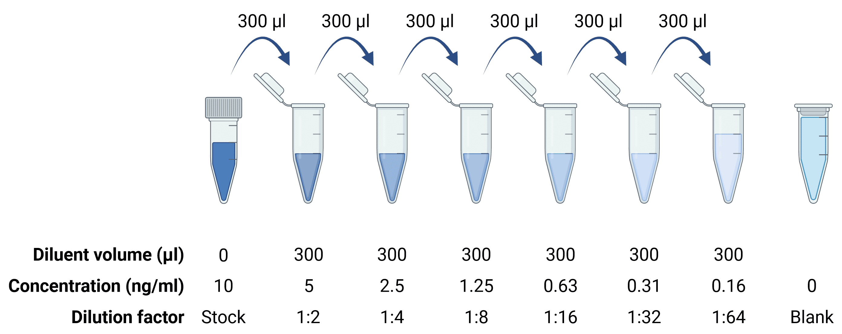
A standard stock solution typically represents a concentration of analyte of 1,000-10,000 pg/ml, which is then serially diluted 6 or 7 times to create the standards needed for the standard curve. For higher sensitivity ELISAs that are capable of detecting antigen to sub-picogram levels, a standard curve will start at a lower stock concentration, but the procedure will be the same, performing serial 1:2 or 1:3 dilutions.

  • Create the stock solution, for example 10,000 pg/ml, by dissolve 10 ng in 1 ml of sample diluent.
  • Add 300 μl to each of 7 other tubes, labeled as follows: 1:2, 1:4, 1:8, 1:16, 1:32, 1:64 and 1:128 to represent the dilution factors.
    • These correspond to 5000, 2500, 1250, 625, 312.5, 156.3, 78.1 pg/ml.
  • Take 300 μl of the stock 10,000 pg/ml solution and add it to the 300 μl in the 1:2 tube. Mix thoroughly.
  • Take 300 μl from the 1:2 tube and add it to the 300 μl in the 1:4 tube. Mix thoroughly.
  • Take 300 μl from the 1:4 tube and add it to the 300 μl in the 1:8 tube. Mix thoroughly.
  • Perform similar dilutions until the full standard range has been created.
    • Standard solutions should be used within approximately 2 hours
  • When ready to perform the ELISA, add 100 μl of each of the standard solutions to the appropriate empty wells. Perform each standard in duplicate or triplicate to ensure accuracy.
Diagram of serial dilutions of a stock standard in 1.5 ml tubes, down to a dilution factor of 1:64

Figure 4: Serial dilutions to generate a standard curve.

All ELISA kits sold by Antibodies.com come with a pre-prepared TMB Substrate solution. To make it from scratch, see the recipes below.

TMB substrate solution

TMB stock solution

  • 10 mg TMB
  • 10 ml DMSO

Phosphate-citrate buffer, 0.5 M

  • 7.1 g Na2HPO4 (anhydrous)
  • 11.5 g citric acid
  • Dissolve in 100 ml distilled water
  • pH 5.6

TMB working chromogen solution

  • 1 ml TMB-DMSO stock
  • 9 ml phosphate-citrate buffer
  • 2 μl H2O2

pNPP substrate solution

0.1M Glycine buffer

  • 751 mg glycine (0.1 M)
  • 20.3 mg MgCl2 (1 mM)
  • 13.6 mg ZnCl2 (1 mM)
  • Dissolve in 100 ml distilled water
  • pH 10.4

Dissolve pNPP in 0.1 M glycine buffer to a concentration of 1 mg/ml.

HRP stop solution

  • 2 M sulfuric acid (H2SO4)

AP stop solution

  • 1 M sodium hydroxide (NaOH)
Side Menu Main Menu Contact Us 0Checkout
Top