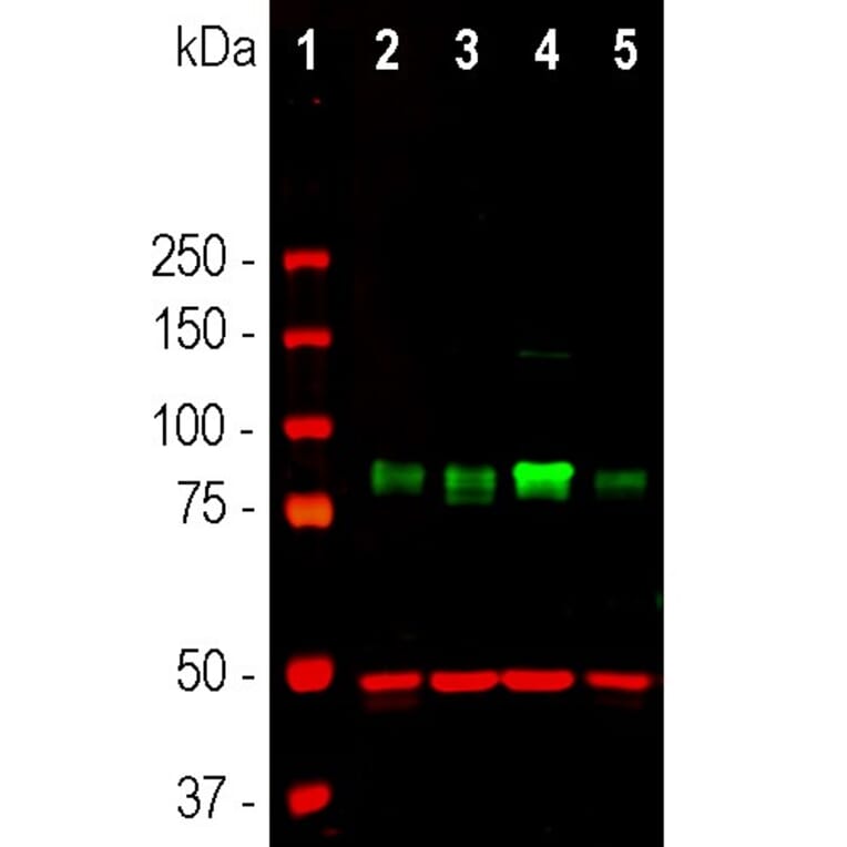
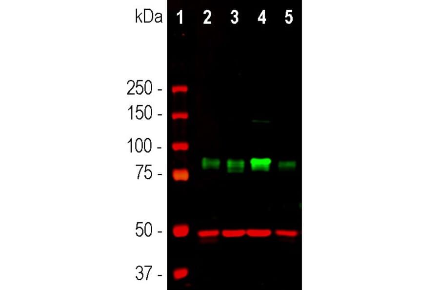
Western blot analysis of different cell lines lysates using Anti-MARCKS Antibody (1:1,000 | green): [1] protein standard (red), [2] HEK293, [3] HeLa, [4] SH-SY5Y, and [5] COS1 cells. Strong band at 80kDa corresponds to MARCKS protein. The same blot was simultaneously probed with Anti-GAPDH Antibody (1:5,000 | red), which reveals a single band at 37kDa in all preparations
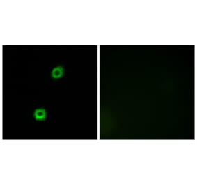
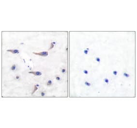
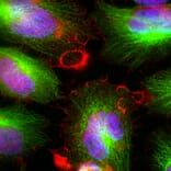
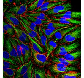
Immunofluorescent analysis of HeLa cells stained with Anti-MARCKS Antibody (1:5,000 ) red, and co-stained with Anti-beta Tubulin Antibody (1:10,000 | green). The blue is Hoechst staining of nuclear DNA. The Anti-MARCKS Antibody binds to MARCKS protein expressed in the plasma membrane and cytoplasm, while the Anti-beta Tubulin Antibody stains cytoplasmic microtubules.
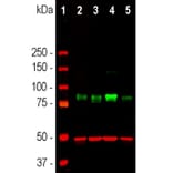
Alternative products to Anti-MARCKS Antibody (A104335)


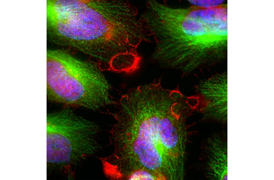
![Immunofluorescence - Anti-MARCKS Antibody [5F9] (A270556) - Antibodies.com](https://cdn.antibodies.com/image/catalog/270/A270556_1.jpg?profile=product_alternative)