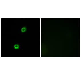
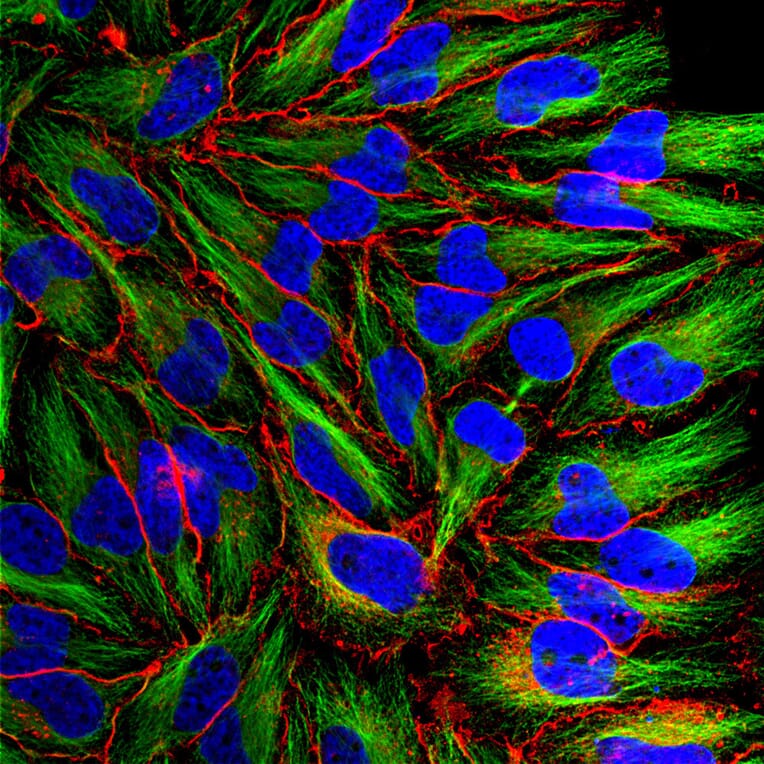
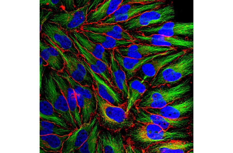
Immunofluorescent analysis of HeLa cells stained with Anti-MARCKS Antibody, at a dilution of 1:1,000, in red, and Anti-Beta Tubulin Antibody (A85428 | 1:10,000, in green. The blue is DAPI staining of nuclear DNA. Anti-MARCKS Antibody recognizes protein expressed in the plasma membrane and cytoplasm, while the Anti-Beta Tubulin Antibody stains microtubules.
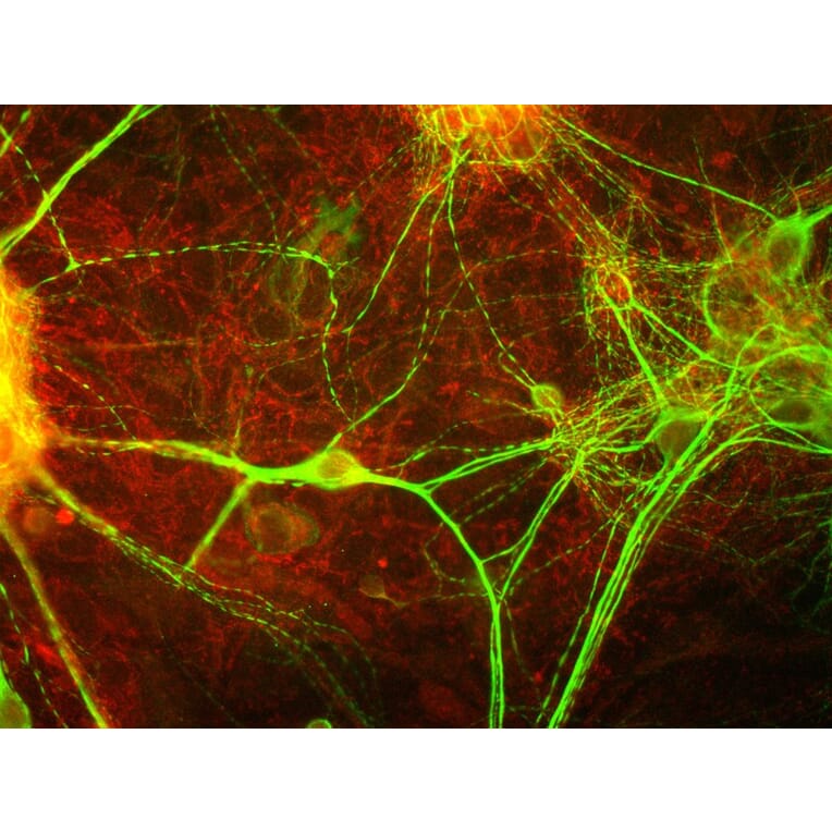
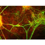
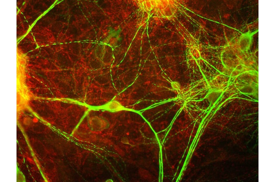
View of mixed neuron/glial cultures stained with Anti-MARCKS Antibody (red) and Anti-MAP2 Antibody (A85363 | green). Note that the Anti-MARCKS Antibody stains vesicular structures both in the glial cells and in the dendrites of the neurons, which are strongly stained with the Anti-MAP2 Antibody.
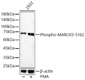
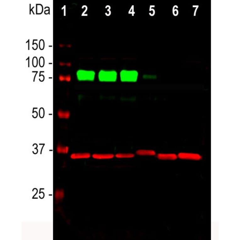
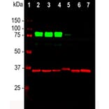
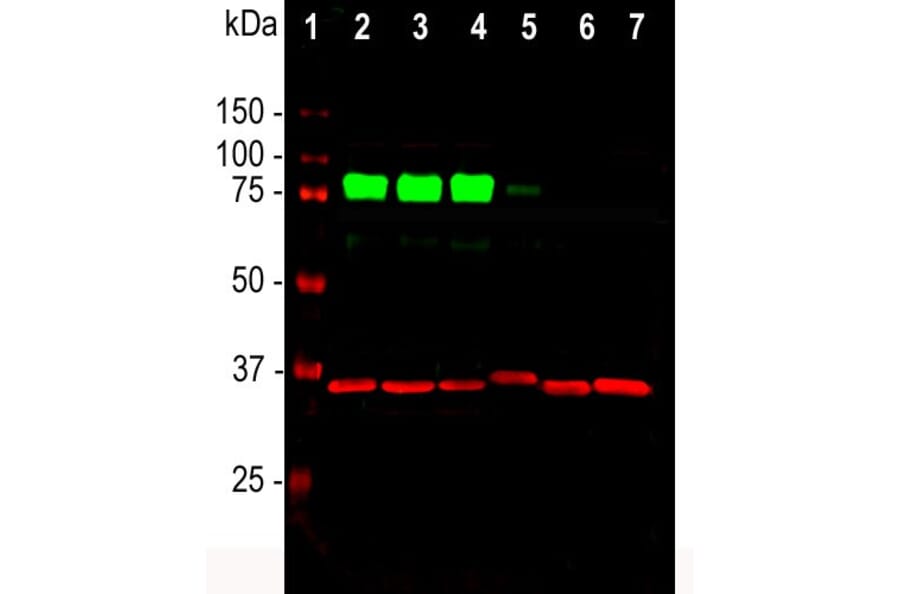
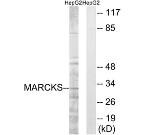
Western blot analysis of cell line lysates probed simultaneously with Anti-MARCKS Antibody, at a dilution of 1:1,000, in green, and Anti-GAPDH Antibody (A85382 | 1:5,000, in red,: [Lane 1] protein standard (red), [Lane 2] HEK293, [Lane 3] HeLa, [Lane 4] SH-SY5Y, [Lane 5] COS1, [Lane 6] NIH-3T3, [Lane 7] C6 cells. The strong band at 80kDa corresponds to MARCKS protein, detected only in the proteins of human origin. Slight reactivity is observed in the monkey cells [Lane 5], but no reactivity is seen on rodent cells. Anti-GAPDH Antibody was used as a loading control - revealing a single band at ~37kDa in all preparations.
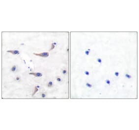
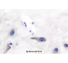
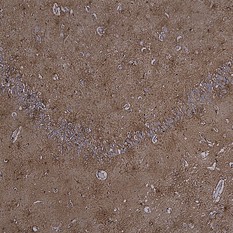
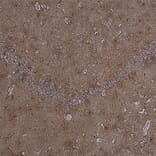
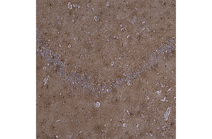
Immunohistochemistry analysis of a formalin fixed paraffin embedded human hippocampus section with Anti-MARCKS Antibody (A85457) at a dilution of 1:2,000. Anti-MARCKS Antibody (A85457) labels the nuclei, membrane and cytoplasm of glial cells and the synapses of neuronal cells. Note: this antibody performs well in the staining of NBF fixed human tissues but does not cross react with rodent.
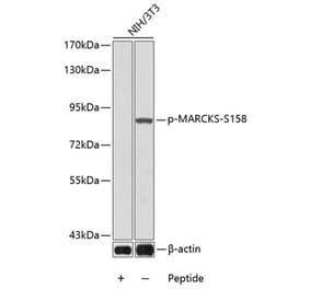
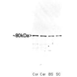
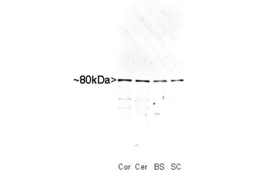
Western blot of whole rat cortex (Co), cerebellum (Ce), brain stem (BS) and spinal cord (SC) homogenate stained with Anti-MARCKS Antibody, at a dilution of 1:10,000). A prominent band running with an apparent SDS-PAGE molecular weight of ~80 kDa corresponds to MARCKS.


![Immunofluorescence - Anti-MARCKS Antibody [5F9] (A270556) - Antibodies.com](https://cdn.antibodies.com/image/catalog/270/A270556_1.jpg?profile=product_alternative)