Human, Mouse, Rat, Beluga, Bovine, Canine, Fish, Carp, Guinea Pig, Hamster, Monkey, Porcine, Sheep, Coral, Plant, Shark, L. amazonensis
Cross Reactivity
This antibody may cross-react with HSC70 at lower dilutions.
Immunogen
Recombinant Full length Human HSP70 Protein.
Host
Rabbit
Clonality
Polyclonal
Isotype
IgG
Conjugate
Unconjugated
Purification
Peptide affinity purification.
Concentration
1 mg/ml
Molecular Weight
~70 kDa
Product Form
Liquid
Formulation
Supplied in Phosphate Buffered Saline, pH 7.4, with 50% Glycerol and 0.09% Sodium Azide.
Storage
Shipped at 4°C. Upon delivery aliquot and store at -20°C. Avoid freeze / thaw cycles.
Synonyms
Heat shock 70 kDa protein 1, Heat shock 70 kDa protein 1A, Heat shock 70 kDa protein 1B, Heat shock 70 kDa protein 2, Heat shock protein family A member 1A, Heat shock protein family A member 1B, HSP70-1, HSP70-2, HSP70.1, HSP70.2, HSP72, HSPA1, HSPA1A, HSPA1B, HSX70
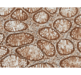
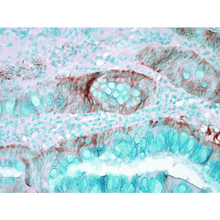
Immunohistochemistry analysis of mouse inflamed colon, fixed in formalin. The Primary Antibody used was Anti-HSP70 Antibody (A304824) at 1:1,000 for 12 hours at 4°C. The secondary antibody used was Biotin Goat Anti-Rabbit at 1:2000 for 1 hour at room temperature. Counterstain: Methyl Green at 200uL for 2 min at room temperature.
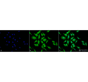
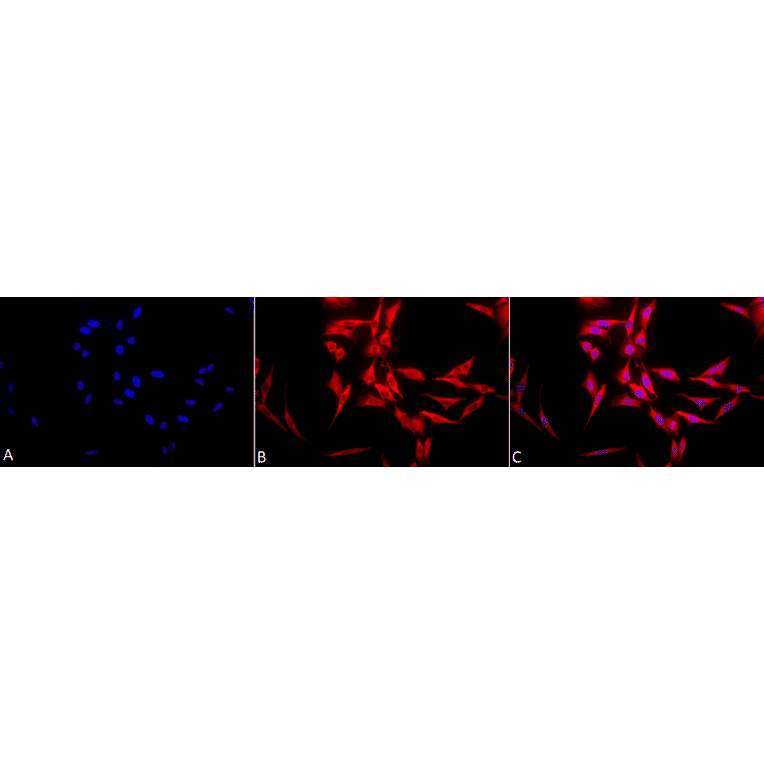
Immunocytochemistry/Immunofluorescence analysis of human heat shocked cervical cancer cell line (HeLa), fixed in 2% formaldehyde for 20 minutes at room temperature, using Anti-HSP70 Antibody (A304824), at 1:100 for 12 hours at 4°C. The secondary antibody used was APC Goat Anti-Rabbit (red) at 1:200 for 2 hours at room temperature. Counterstain: DAPI (blue) nuclear stain at 1:40000 for 2 hours at room temperature. Localization: Cytoplasm. Magnification: 20x.(A) DAPI (blue) nuclear stain. (B) Anti-Hsp70 Antibody. (C) Composite. Heat Shocked at 42°C for 1h.
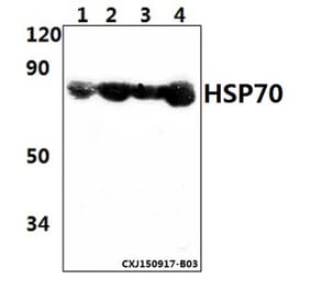
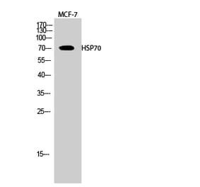
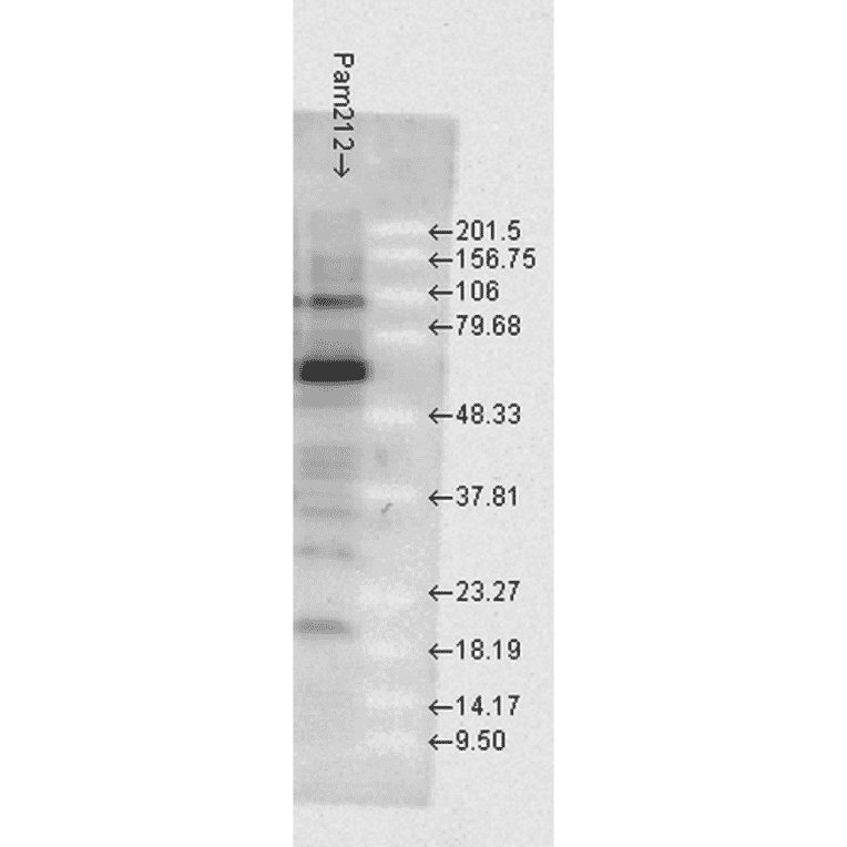
Western blot analysis of mouse Pam212 cells showing detection of HSP70 protein using Anti-HSP70 Antibody (A304824) at 1:1,000 for 2 hours at room temperature. Load: 15 µgprotein. Block: 1.5% BSA for 30 minutes at room temperature. The secondary antibody used was Donkey Anti-Rabbit IgG: HRP for 1 hour at room temperature.
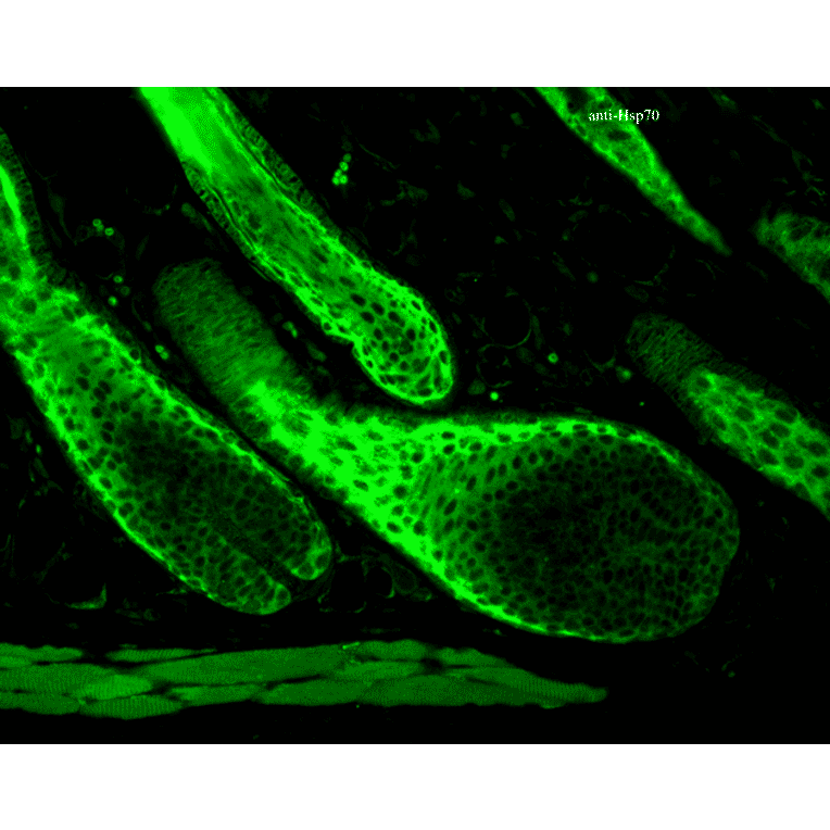
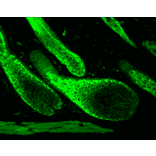
Immunohistochemistry analysis of mouse backskin, fixed in Bouin's fixative solution. The Primary Antibody used was Anti-HSP70 Antibody (A304824) at 1:100 for 1 hour at room temperature. The secondary antibody used was FITC Goat Anti-Rabbit (green) at 1:50 for 1 hour at room temperature. Localization: Cytoplasm.
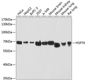
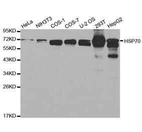
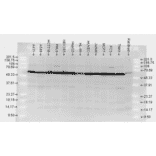
Western blot analysis of Human, rat brain cell lysates showing detection of HSP70 protein using Anti-HSP70 Antibody (A304824) at 1:10,000 for 2 hours at room temperature. Load: 2 µg. Block: 1.5% BSA for 30 minutes at room temperature. The secondary antibody used was Donkey Anti-Rabbit IgG: HRP for 1 hour at room temperature.
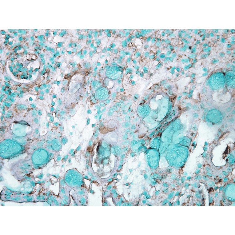
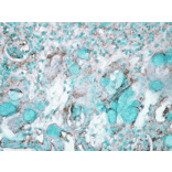
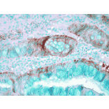
Immunohistochemistry analysis of human colon carcinoma, fixed in formalin. The Primary Antibody used was Anti-HSP70 Antibody (A304824) at 1:50,000 for 12 hours at 4°C. The secondary antibody used was Biotin Goat Anti-Rabbit at 1:2000 for 1 hour at room temperature. Counterstain: Methyl Green at 200uL for 2 min at room temperature.
Immunocytochemistry/Immunofluorescence analysis of human heat shocked cervical cancer cell line (HeLa), fixed in 2% formaldehyde for 20 minutes at room temperature, using Anti-HSP70 Antibody (A304824), at 1:100 for 12 hours at 4°C. The secondary antibody used was FITC Goat Anti-Rabbit (green) at 1:200 for 2 hours at room temperature. Counterstain: DAPI (blue) nuclear stain at 1:40000 for 2 hours at room temperature. Localization: Cytoplasm. Magnification: 100x.(A) DAPI (blue) nuclear stain. (B) Anti-Hsp70 Antibody. (C) Composite. Heat Shocked at 42°C for 1h.


![FACS - Anti-HSP70 Antibody [C92F3A-5] (A304773) - Antibodies.com](https://cdn.antibodies.com/image/catalog/304/A304773_1.png?profile=product_alternative)
![Western Blot - Anti-HSP70 Antibody [N27F3-4] (A305087) - Antibodies.com](https://cdn.antibodies.com/image/catalog/305/A305087_1.png?profile=product_alternative)
![Immunohistochemistry - Anti-HSP70 Antibody [BB70] (A305113) - Antibodies.com](https://cdn.antibodies.com/image/catalog/305/A305113_1.png?profile=product_alternative)
![Western Blot - Anti-HSP70 Antibody [3A3] (A305077) - Antibodies.com](https://cdn.antibodies.com/image/catalog/305/A305077_1.png?profile=product_alternative)
![Western Blot - Anti-HSP70 Antibody [5A5] (A305076) - Antibodies.com](https://cdn.antibodies.com/image/catalog/305/A305076_1.png?profile=product_alternative)
![FACS - Anti-HSP70 Antibody [1H11] (A304712) - Antibodies.com](https://cdn.antibodies.com/image/catalog/304/A304712_1.png?profile=product_alternative)