Human Rab5 synthetic peptide conjugated to KLH; identical to dog Rab5 sequence over the residues.
Hôte
Rabbit
Clonalité
Polyclonal
Isotype
IgG
Conjuguer
Unconjugated
Purification
Protein A purification.
Concentration
1 mg/ml
Masse moléculaire
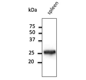
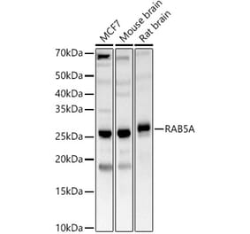
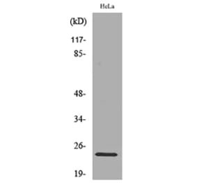
~26 kDa
Forme du produit
Liquid
Formulation
Supplied in Phosphate Buffered Saline with 50% Glycerol and 0.09% Sodium Azide.
Stockage
Shipped at 4°C. Upon delivery aliquot and store at -20°C. Avoid freeze / thaw cycles.
Synonymes
RAB 5, RAB 5A, RAB5A, RAB5A member RAS oncogene family, RAB5A_HUMAN, RAS associated protein RAB5A, Ras related protein Rab 5A, Ras-related protein Rab-5A
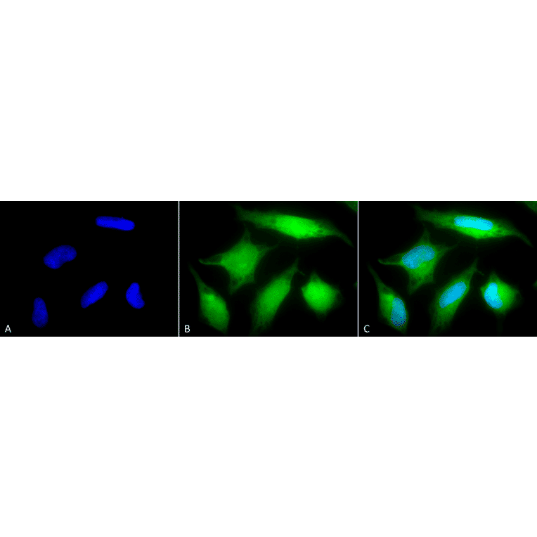
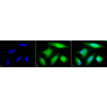
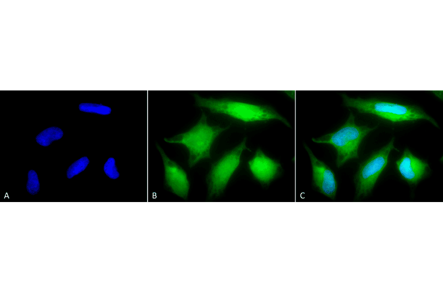
Immunocytochemistry/Immunofluorescence analysis of human cervical cancer cell line (HeLa), fixed in 2% formaldehyde for 20 minutes at room temperature, using Anti-Rab5 Antibody (A305104), at 1:80 for 12 hours at 4°C. The secondary antibody used was FITC Goat Anti-Rabbit (green) at 1:200 for 2 hours at room temperature. Counterstain: DAPI (blue) nuclear stain at 1:40000 for 2 hours at room temperature. Localization: Cytoplasm. Melanosome. Nucleus. Magnification: 20x.(A) DAPI (blue) nuclear stain. (B) Anti-Rab5 Antibody. (C) Composite.
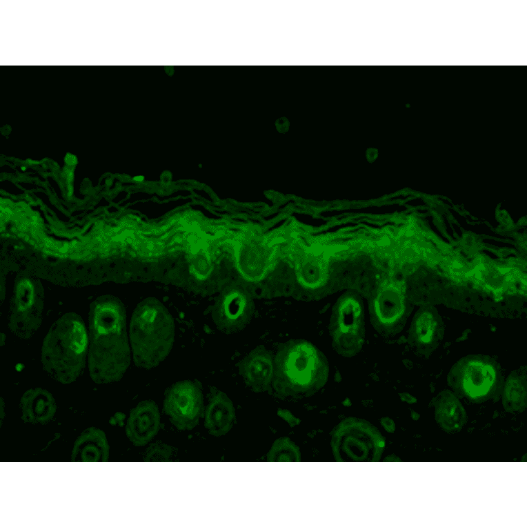
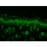
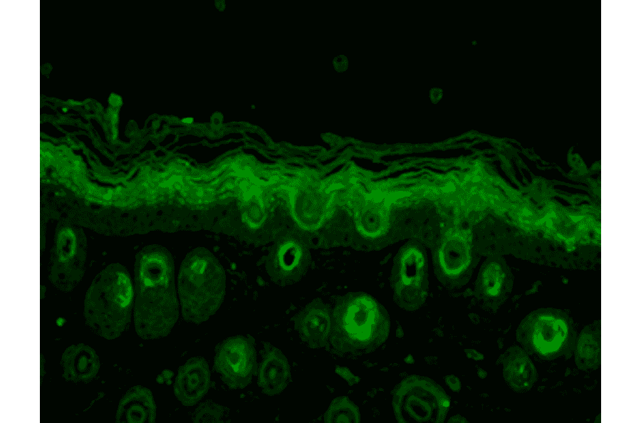
Immunohistochemistry analysis of mouse backskin, fixed in Bouin's fixative solution. The Primary Antibody used was Anti-Rab5 Antibody (A305104) at 1:100 for 1 hour at room temperature. The secondary antibody used was FITC Goat Anti-Rabbit (green) at 1:50 for 1 hour at room temperature. Localization: Cytoplasm.
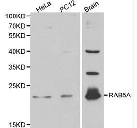
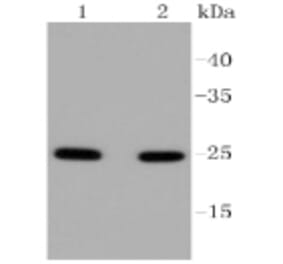
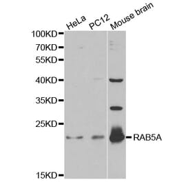
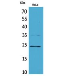
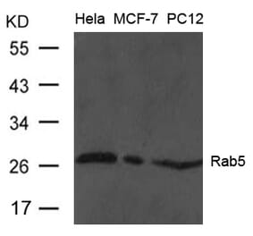
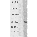
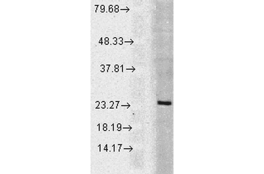
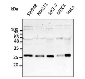
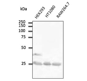
Figure 3: Western Blot - Anti-Rab5 Antibody (A305104)
Western blot analysis of human Cell line lysates showing detection of Rab5 protein using Anti-Rab5 Antibody (A305104) at 1:1,000 for 2 hours at room temperature. Load: 15 µgprotein. Block: 1.5% BSA for 30 minutes at room temperature. The secondary antibody used was Donkey Anti-Rabbit IgG: HRP for 1 hour at room temperature.
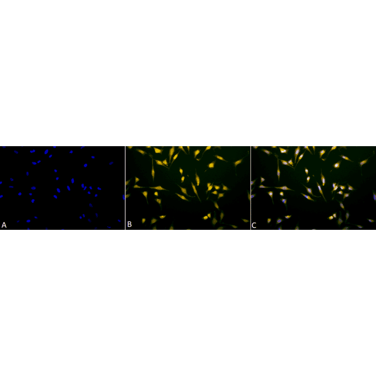
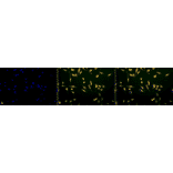
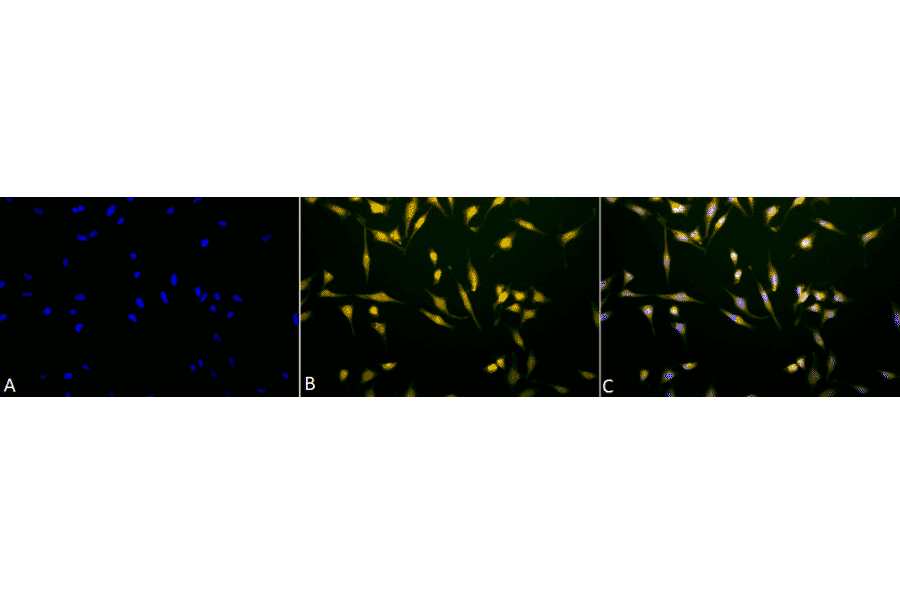
Immunocytochemistry/Immunofluorescence analysis of human cervical cancer cell line (HeLa), fixed in 2% formaldehyde for 20 minutes at room temperature, using Anti-Rab5 Antibody (A305104), at 1:80 for 12 hours at 4°C. The secondary antibody used was R-PE Goat Anti-Rabbit (yellow) at 1:200 for 2 hours at room temperature. Counterstain: DAPI (blue) nuclear stain at 1:40000 for 2 hours at room temperature. Localization: Cytoplasm. Melanosome. Nucleus. Magnification: 100x.(A) DAPI (blue) nuclear stain. (B) Anti-Rab5 Antibody. (C) Composite.
Produits alternatifs à Anti-Rab5 Anticorps (A305104)


![Western Blot - Anti-Rab5A Antibody [ARC0297] (A306380) - Antibodies.com](https://cdn.antibodies.com/image/catalog/306/A306380_1.jpg?profile=product_alternative)