

Ryan Hamnett, PhD | 25th January 2025
Neurons are electrically excitable cells in the nervous system that sense, process and transmit information to control the actions of the body. Pan-neuronal markers are proteins that are expressed across all or most neurons due to their importance to fundamental neuronal functions such as synaptic transmission.
Pan-neuronal markers can be detected using specific antibodies in techniques such as immunohistochemistry (IHC) and immunocytochemistry/immunofluorescence (ICC/IF). Marking out all neurons is important for determining whether a protein of interest is expressed in neurons, and for monitoring how mature neuron numbers change during development, neurogenesis, and neurodegenerative conditions such as Alzheimer’s disease.

Neurons are highly compartmentalized cells, including the cell body (or soma), axons, dendrites, and synapses. This guide classifies pan-neuronal markers based on their subcellular localization; different markers highlight different morphological features in neurons, so the choice of pan-neuronal marker will depend on what the experiment aims to see.
It is important to note that no ‘pan-neuronal’ marker truly marks every neuronal subtype. For instance, some neurons in the hypothalamus and cerebellum are known not to express NeuN, while other pan-neuronal markers, such as PGP9.5 or HuC/D, may be pan-neuronal in peripheral neurons but not the central nervous system.
Pan-neuronal markers that highlight the nucleus or cell body are ideal for counting the numbers of neurons, without neuronal fibers complicating the picture.
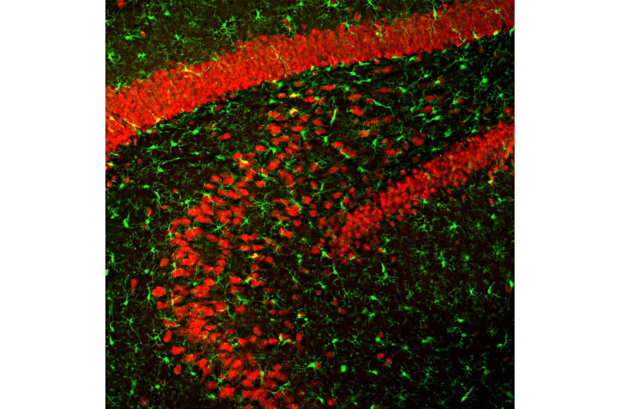
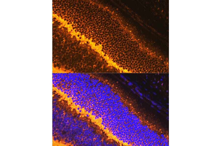
NeuN, also known as Fox-3, is the most commonly used marker of neuronal nuclei. A regulator of RNA splicing, NeuN is strongly expressed in the nuclei of most neurons of the central nervous system and is absent from other cell types such as glia.1 Not all neurons are positive for NeuN, with some neurons of the cerebellum (e.g. Purkinje cells), retina, spinal cord and suprachiasmatic nucleus lacking NeuN expression.1,2
The neuron-specific RNA-binding proteins HuC and HuD are the products of the ELAVL3 and ELAVL4 genes. Pan-specific antibodies against Hu proteins have been shown to be pan-neuronal markers in the brain, particularly earlier in development, while antibodies that bind to HuC/D highlight the nucleus and soma of all neurons in the enteric nervous system.3,4
PGP9.5, also known as UCHL1, is a highly conserved and neuron-specific ubiquitin hydrolase enzyme. PGP9.5 is localized to the cytoplasm, including the cell body and neuronal processes, and has been estimated to constitute as much as 10% of total cytoplasmic protein in neurons.5 As such, it is useful for highlighting the complete neural network of organs.5
Neuron-specific enolase (NSE) is a cytoplasmic marker of neurons and neuroendocrine cells. Because elevated levels of NSE in the serum have proven useful for medical diagnoses, NSE is most commonly used as a biomarker of CNS insults such as stroke and ischemia, or for cancer, particularly small cell lung cancer, non-small cell lung cancer, neuroendocrine tumors, and neuroblastoma.6
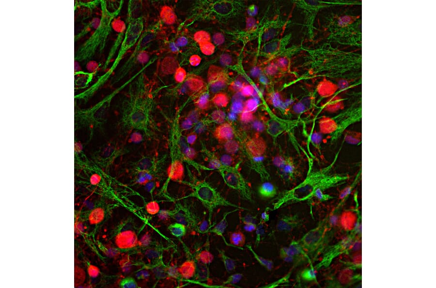
Figure 3: IF of cortical neuron-glial cell culture from E20 rat stained with chicken anti-UCHL1 (A85349) in red and mouse anti-Vimentin (A85423) in green. Nuclei are stained blue with DAPI.
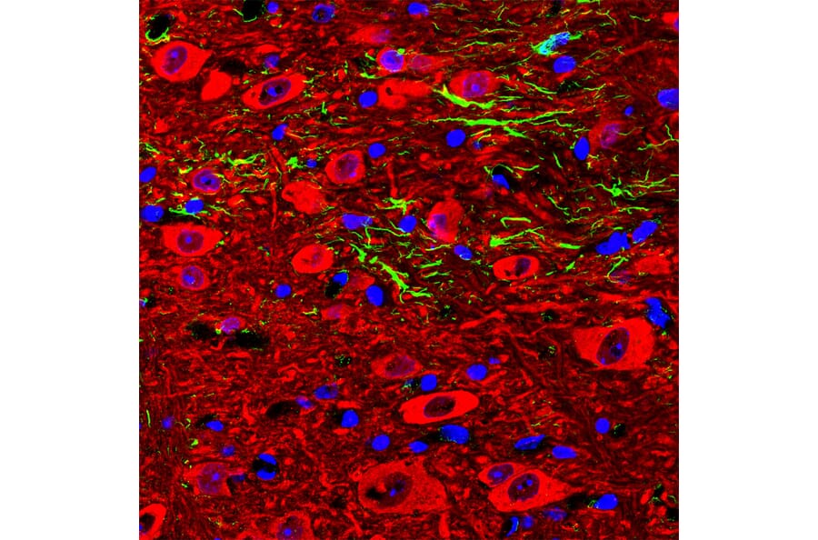
Figure 4: IHC of a section of adult mouse cerebellar dentate nucleus stained with chicken anti-NSE (A104334) in red and anti-GFAP in green. Nuclei are stained blue with Hoechst.
Pan-neuronal markers that can stain neuronal processes are useful for investigating neuronal interactions with other neurons. This is particularly useful in the peripheral nervous system, where neurons directly project to and innervate organs and muscles, during development to identify mature neurons, and in vitro in neuronal cell culture. Markers of axons and dendrites are often cytoskeletal components, which have important roles in neurons in axonal growth and transport of components along the length of neuronal processes.
Beta III tubulin (βIII-tubulin) is a microtubule-associated protein found almost exclusively in neurons. βIII-tubulin is found in mature neurons, but also during neurogenesis in developing neurons.7 MAP2 is a microtubule-associated protein involved in microtubule stabilization that is frequently used as a pan-neuronal marker. Unlike beta III tubulin, MAP2 is found in dendrites and soma but not the axon, making it useful for establishing the direction of travel for information between neurons.8 MAP2 is closely related to the MAP tau (MAPT) protein, sometimes known as simply tau, which marks dendrites, perikarya and axons. While MAPT is a well-established neuron marker, it is best known for its involvement in the development of Alzheimer’s disease and related tauopathies. In affected brains, tau becomes hyperphosphorylated and clumps together in neurofibrillary tangles, disrupting normal neuronal function and triggering neurodegeneration.9
Figure 5: IF of cortical neuron-glial culture from E20 rat stained with mouse anti-Tau (A85416) in green and chicken anti-MAP2 (A85363) in red. Nuclei are stained blue with DAPI.
Figure 6: IF of P19 mouse embryonal carcinoma cells stimulated to neuronal differentiation by retinoic acid, stained with mouse anti-Beta III Tubulin [TU-20] (A86691) in red. Nuclei are stained blue with DAPI.
Finally, the light, medium and heavy neurofilament proteins NF-L, NF-M and NF-H are neuron-specific intermediated filament proteins. Named for their respective molecular masses of approximately 70, 160 and 200 kDa, NF-L, NF-M and NF-H are highly abundant in axons, where they provide structural support for long projections.10 As well as being biomarkers in normal health, neurofilament levels in cerebrospinal fluid (CSF) has been found to increase in some neurodegenerative conditions such as multiple sclerosis.11 Peripherin is an additional neurofilament protein that is used as a pan-neuronal marker primarily in the peripheral nervous system.
While some proteins involved in synaptic transmission, such as VGLUT1/2 in glutamatergic neurons and vAChT in cholinergic neurons, are found only in specific subtypes, many synaptic proteins are found in the majority of neurons as part of the basic machinery of neurotransmission. Synaptic markers are ideal for generating high resolution images of neuronal interactions, but many are found only at dendritic spines or axon terminals, providing no information about neuronal morphology or even which neuron they belong to. For that reason, they are often stained alongside cytoplasmic markers such as MAP2 or fluorescent reporters.
Pre-synaptic proteins involved in the fusion of synaptic vesicles to the plasma membrane include synapsin, SNAP25, synaptotagmin, synaptobrevin (VAMP2) and synaptophysin, which regulate the release of neurotransmitters into the synaptic cleft. Post-synaptic proteins that provide structure and support to dendritic spines include members of the DLG and SHANK families. PSD95, also known as DLG4, organizes receptors and channels on the post-synaptic membrane. Other post-synaptic proteins include neurotransmitter receptors, but because these are neurotransmitter-specific, they cannot be considered pan-neuronal.
![IHC of rat brainstem using anti-NeuN [1B7] (A85405)](https://cdn.antibodies.com/image/catalog/85/A85405_4.jpg?profile=product_image)

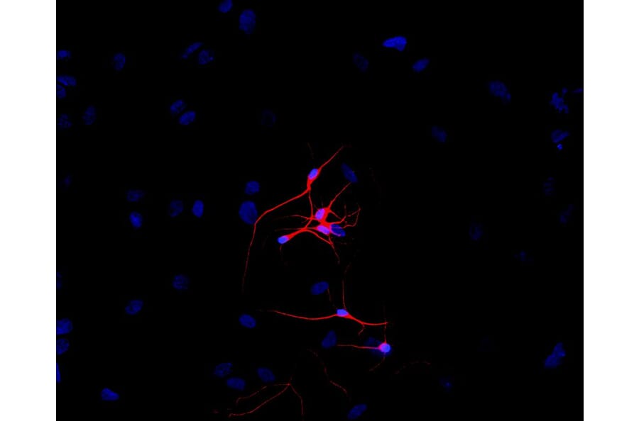


![ICC/IF of rat hippocampal neurons using Anti-PSD95 [6G6] Antibody (A304703)](https://cdn.antibodies.com/image/catalog/304/A304703_2.png?profile=product_image)