Ryan Hamnett, PhD | 12th June 2024
Loading controls are one of the most important controls to run in a western blot, allowing researchers to identify variation introduced as a result of unequal protein extraction, uneven protein loading, inconsistent transfer from gel to membrane, or patchy signal detection.
A loading control effectively acts as an internal reference protein, allowing researchers to ensure that differences observed between samples are due to genuine biological variation rather than experimental error.
The protein chosen for a loading control should not vary with experimental conditions, meaning any difference seen in the levels of this protein actually reflect procedural error. Not only does this identify unintended variation, it also enables error correction by normalizing the signal of the protein of interest to the loading control signal.
Checking that total protein concentration is consistent across samples is essential to know whether differences between samples are biological or not. Loading controls are used as a proxy for total protein content. Earlier steps in the western blotting procedure, such as a BCA, Bradford or Lowry assay, aim to keep protein content consistent prior when loading samples on to the gel. Loading controls check that loading was equal, in addition to identifying potential issues with membrane transfer and signal detection, therefore allowing the examination of biological differences between samples (Figure 1). Both loading controls and early protein quantification are recommended when performing a western blot for the most reliable and consistent results.
Figure 1: Beta-actin loading control validating a KO experiment. Western blot using anti-Cyclin D1 antibody in WT and Cyclin D1 knockout HeLa cells. No band is observed for cyclin D1 in the right-hand lane, but total protein levels are unaffected as confirmed by the beta Actin loading control. Therefore, the knockout of Cyclin D1 has been successfully verified.
Loading control proteins tend to be housekeeping genes: well-understood and ubiquitously expressed proteins that are involved in the basic functioning of the cell. They are therefore unlikely to vary between cell types or to be affected by experimental treatments, though this must be verified to prevent incorrect normalization of genuine biological variation.
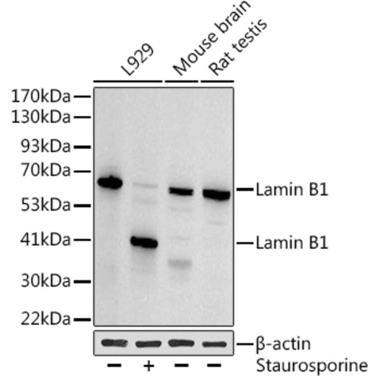
Figure 2: Loading controls can be affected by experimental conditions.Western blot using anti-Lamin B1 antibody, which can act as a nuclear loading control. However, lamin B1 is cleaved during apoptosis3, caused here by staurosporine addition, which would make it unsuitable for all experiments. Instead, beta Actin is used as a loading control.
| Subcellular localization / Sample type | Control name | Molecular weight, kDa |
|---|---|---|
| Whole cell/cytoplasmic | alpha-Tubulin | 55 |
| beta-Actin | 43 | |
| beta-Tubulin | 55 | |
| Cofilin | 19 | |
| Cyclophilin A | 18 | |
| Cyclophilin B | 18 | |
| GAPDH | 37 | |
| Vinculin | 116 | |
| Nuclear | HDAC1 | 62 |
| Histone H1 | 15 | |
| Histone H2B | 15 | |
| Histone H3 | 15 | |
| Lamin B1 | 66 | |
| PCNA | 29 | |
| TATA binding protein (TBP) | 38 | |
| Mitochondrial | COX IV | 17 |
| Hsp60 | 60 | |
| VDCA1 | 31 | |
| Plasma membrane | beta-Catenin | 86 |
| CD44 | 82 | |
| Na+/K+ ATPase alpha-1 | 112 | |
| Muscle | SDHA | 73 |
| Serum | Transferrin | 77 |
Table 1:Loading controls to use in western blot experiments
| Target | Antibody | Reactivity |
|---|---|---|
| Sodium Potassium ATPase | Anti-ATPase Antibody (A95104) | Human, Mouse, Rat |
| Target | Antibody | Reactivity |
|---|---|---|
| COX IV | Anti-COX IV Antibody (A82550) | Human |
| Anti-COX IV Antibody (A9916) | Human, Mouse, Rat | |
| Heat Shock Protein 60 | Anti-Heat Shock Protein 60 Antibody (A85438) | Human, Mouse, Rat |
| Anti-Heat Shock Protein 60 Antibody (A85437) | Human, Mouse, Rat | |
| Anti-Heat Shock Protein 60 Antibody [1C7] (A85439) | Human, Mouse, Rat |
| Target | Antibody | Reactivity |
|---|---|---|
| HDAC1 | Anti-HDAC1 Antibody (A12564) | Human, Mouse, Rat |
| Anti-HDAC1 Antibody (A84192) | Human, Mouse | |
| Histone H3 | Anti-Histone H3 Antibody (A25203) | Human, Mouse, Rat |
| Anti-Histone H3 Antibody (A16702) | Human, Mouse, Rat | |
| Anti-Histone H3 Antibody [RM186] (A121358) | All Vertebrates | |
| Anti-Histone H3 Antibody [RM190] (A121350) | All Vertebrates | |
| Lamin B1 | Anti-Lamin B1 Antibody (A82838) | Human, Mouse |
| Anti-Lamin B1 Antibody (A26974) | Human, Mouse, Rat | |
| PCNA | Anti-PCNA Antibody (A83563) | Human, Mouse, Rat, Porcine |
| Anti-PCNA Antibody (A25315) | Human, Mouse, Rat | |
| Anti-PCNA Antibody [PC10] (A86878) | Human, Mouse, Rat, Chicken, Drosophila | |
| TBP | Anti-PCNA Antibody (A26155) | Human, Mouse, Rat |
| Anti-PCNA Antibody (A82888) | Human, Mouse, Rat |
| Target | Antibody | Reactivity |
|---|---|---|
| Transferrin | Anti-Transferrin Antibody [HTF-14] (A85619) | Human, Rabbit, Porcine |
| Anti-Transferrin Antibody [PTF-02] (A85822) | Porcine |
| Target | Antibody | Reactivity |
|---|---|---|
| Cyclophilin B | Anti-Cyclophilin B Antibody [Pk2E2AT] (A58461) | Human |
| GAPDH | Anti-GAPDH Antibody (A83722) | Human, Mouse, Rat |
| Anti-GAPDH Antibody (A85377) | Human, Horse, Cow, Porcine, Chicken, Rat, Mouse | |
| Anti-GAPDH Antibody [1D4] (A85382) | Human, Horse, Cow, Porcine, Chicken, Rat, Mouse | |
| Anti-GAPDH Antibody [GA1R] (A85271) | Chicken, Hamster, Human, Mouse, Rat, Rabbit, Sf9 Insect, BL-21 Bacteria, S. cerevisiae | |
| Anti-GAPDH Antibody [RM114] (A121396) | Human, Monkey | |
| Vinculin | Anti-Vinculin Antibody (A94871) | Human, Mouse, Rat |
![Western blot witb beta-Actin loading control - Anti-Cyclin D1 Antibody [ARC0300] (A306339) - Antibodies.com](https://cdn.antibodies.com/image/catalog/306/A306339_1.jpg?profile=product_top)