Oligodendrocyte Markers

Kai Boon Tan, PhD | 22nd August 2025

Oligodendrocytes are a specialized type of glial cell in the central nervous system (CNS) that produces myelin, a fatty insulating substance that wraps around neuronal axons. This myelin sheath serves to speed up the transmission of electrical impulses along nerve fibers, thereby enabling rapid and efficient communication between neurons.

Oligodendrocyte generation, maturation, and functional diversity are orchestrated by a tightly regulated sequence of transcriptional programs and signaling events. Here, we describe oligodendrocyte lineage progression, maturation, functional roles in myelination and remyelination, and compile common molecular markers at each stage.

Oligodendrocyte markers are important tools in neuroscience research. Below are some examples of how oligodendrocyte markers can be used in research:

  • Study the roles of oligodendrocytes in myelination and remyelination dynamics in development and brain injury.
  • Interrogate cell fate specification in oligodendrocytes in combination with genetic fate-mapping approaches.
  • Isolate specific oligodendrocyte or oligodendrocyte progenitor cell (OPC) populations using fluorescence-activated cell sorting (FACS), enabling downstream procedures such as in vitro differentiation, transplantation, or molecular profiling.
  • Visualize the heterogeneity of the oligodendrocyte populations to complement single-cell or spatial transcriptomic assays.

Table of Contents

Oligodendrocyte Marker Summary

OPC or oligodendrocyte stage Key Marker(s) Description
Pan-oligodendroglia lineage OLIG2, SOX10 OLIG2 activates SOX10 expression, and their co-expression is active throughout the oligodendrocyte development, differentiation and maturation
OPCs NKX2.2, PDGFRα, NG2 (CSPG4), CXCR4 Transcription factors responsible for OPC fate commitment, proliferation, survival and migration
Early Differentiation MYRF Transcription factors to activate myelin genes
Pre-Myelinating Oligodendrocytes O4, BCAS1 Markers of newly differentiating oligodendrocytes
Mature Myelinating Oligodendrocytes MBP, PLP1, CNPase, MOG, CC1 Structural proteins localize to compact and non-compact myelin
Remyelinating Oligodendrocytes GSTπ, ASPA Marker for newly differentiated oligodendrocytes in response to injury
Mature Oligodendrocytes MCT1 Metabolic support for axons via lactate transport
IHC analysis using Anti-Myelin Basic Protein Antibody (A85321)

Figure 1: Immunofluorescence of rat brain cerebellum stained with Anti-Myelin Basic Protein Antibody [7D2] (A85329) (red) and Anti-NF-M Antibody (A85323) (green). Nuclei are marked by DAPI in blue.

 Immunofluorescence of rat cerebellum section stained with Anti-CNPase Antibody [1H10] (A85413) in green and Anti-NF-M Antibody (A85324) in red.

Figure 2: IHC of mouse brain stained with Anti-Myelin PLP Antibody (A306001) in red. Nuclei were stained by DAPI in blue.

Origin and Specification of Oligodendrocyte Progenitor Cells (OPCs)

Oligodendrocytes are derived from oligodendrocyte progenitor cells, which originate from radial glial cells (RGCs), the primary neural stem cells (NSCs) of the embryonic central nervous system (CNS). As described in the NSC markers page, RGCs line the ventricular zone of the neural tube and generate all major CNS cell types, including neurons, astrocytes, ependymal cells, and oligodendrocytes.

Brain OPC Development

In the forebrain, oligodendrocytes are generated in three waves from the spatiotemporally distinct cohorts of NSCs (Figure 3).

  1. Upon the neural tube closure (see Neural Stem Cell Markers for more information on neurulation), the notochord and floor plate release Sonic Hedgehog (SHH) into the adjacent ventral region of the neural tube. In the forebrain, spatially graded SHH causes RGCs in the medial ganglionic eminence and pre-optic areas to express NKX2.1.2 These NKX2.1+ RGCs later upregulate OLIG1/2 and NKX2.2, thereby committing to oligodendroglia lineage while repressing other neuronal fates (Figure 4).3 As the newly specified OPCs delaminate from the ventricular zone, they express the surface receptors PDGFRα and NG2, which drive OPC proliferation and the migration into the dorsal forebrain, the primitive cerebral cortex, contributing to the first wave of oligodendrocyte generation.2,4,5
  2. Around E15.5 in the mouse, the second wave of the OPCs is generated from GSX2+ cells in the lateral ganglionic eminence. Under the control of GSX2, these OPCs migrate dorsally and later populate the entire forebrain.6
  3. Closer to birth in the mouse, EMX1+ RGCs in the dorsal subventricular zone (SVZ), subjacent to the corpus callosum, generate the third wave of OPCs under the control of SHH released from migratory GABAergic inhibitory interneurons.7–9 These OPCs also express canonical markers such as OLIG2, SOX10, PDGFRα, and NG2, and migrate into the corpus callosum.7,9 By postnatal day (P)10, dorsally derived OPCs dominate callosal regions, effectively outnumbering earlier ventral‑derived populations.7
Schematic representation of the origins of distinct waves of forebrain and spinal cord oligodendrocytes.

Figure 3: Schematic representation of the origins of distinct waves of forebrain and spinal cord oligodendrocytes. At E12.5 in mice, the first wave of forebrain and spinal cord oligodendrocyte precursors (OPCs) is generated from the ventral telencephalon and ventral neural tube. At E15.5, forebrain OPCs arise from the lateral ganglionic eminence, while spinal cord OPCs emerge from the dorsal neural tube. The final wave of forebrain and spinal cord oligodendrogenesis takes place after birth (P0), with forebrain oligodendrocytes produced from dorsal telencephalic progenitors, and spinal cord oligodendrocytes arising from progenitors from the central canal subependymal zone. Edited and reproduced under Creative Commons CC-BY 4.0 from van Tilborg, E. et al. 1

Figure 4: IHC analysis of postnatal mouse brain at postnatal day (P)14 using anti-OLIG1 antibody in blue, NKX2.2 in green, and V5-tagged Tensin3 in red. Edited and reproduced under Creative Commons CC-BY 4.0 from.10

Spinal Cord OPC Development

Similar to the forebrain, spinal cord OPCs originate from the in three distinct NSCs waves in the neural tube:

  1. In the primitive spinal cord, SHH induces oligodendroglia fate by upregulating NKX6.1/6.2, which in turn activate OLIG1/2 in RGCs within the pMN domain.11,12 Like the forebrain OPCs, the first wave of spinal cord OPCs also express PDGFRα and NG2, which enable rapid proliferation and lateral migration along radial glia into ventral white matter (Figure 5). In contrast to the OPCs in the forebrain, OPC differentiation from RGCs in the primitive spinal cord does not require NKX2.1.
  2. The second cohort of OPCs arises from DBX1⁺/ASCL1⁺ progenitors in the dorsal neural tube, independent of SHH. Their specification requires FGF signaling coupled with reduced BMP activity.13,14 These dorsal‑derived OPCs also express PDGFRα and NG2, and preferentially populate the dorsolateral white matter, where they account for < 20% of the total OPC pool. 11,13,15
  3. A final wave emerges around birth from the subependymal zone adjacent to the central canal.16,17 Like earlier waves, these perinatal OPCs maintain PDGFRα+/NG2+ expression, and later downregulate these receptors as they differentiate into myelinating oligodendrocytes.
  4. Figure 5: IHC analysis of mouse adult spinal cord using anti-GFP antibody in green to report ASCL1, (left panel) anti-PDGFRα antibody in red, anti-OLIG2 in blue, and (right panel) anti-NG2 antibody in red. Edited and reproduced under Creative Commons CC-BY 4.0 from Kelenis, D. P. et al.18

Early Differentiation into Oligodendrocytes

As CNS development progresses into late embryonic and early postnatal development, OPCs exit the cell cycle and begin to differentiate into oligodendrocytes. During this process, OPCs maintain OLIG2 expression, which in turn activates SOX10.

anti-SOX10 polyclonal antibody (A90639)

Figure 6: IHC analysis of C6 cells using rabbit Anti-SOX10 polyclonal antibody (A90639) in red and DAPI in blue.

Anti-OLIG2 Antibody [OLIG2/2400] (A248077)

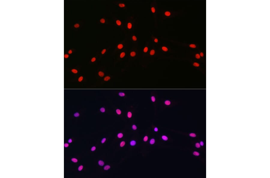
Figure 7: IHC analysis human cerebellum stained with Anti-OLIG2 Antibody [OLIG2/2400] (A248077) in red and DAPI in blue.

Subsequently, SOX10 directly cooperates with Myelin Regulatory Factor (MYRF) to suppress progenitor genes such as PDGFRA and NG2, while activating expression of myelin proteins, including Myelin Basic Protein (MBP), Proteolipid Protein 1 (PLP1), and Myelin Oligodendrocyte Glycoprotein (MOG) to prepare for myelination.19–21 Early during this transition, newly differentiated oligodendrocytes start to express the sulfatide epitope O4, on the cell surface and the cytoplasmic protein BCAS1, both of which are essential in the oligodendrocyte terminal differentiation from OPCs (Figure 8).22,23

Figure 8: IHC analysis of oligodendroglia cell culture using anti-BCAS1 antibody in magenta, anti-MBP antibody in red, anti-Thioflavin S antibody in green, and DAPI in blue. Edited and reproduced under Creative Commons CC-BY 4.0 from Kaji, S. et al.24

Myelination and Neuronal Support by Mature Oligodendrocytes

Peak myelination takes place in the early postnatal weeks in rodents25,26 but extends through adolescence, with remodeling myelination persisting into adulthood in humans.27,28 During peak myelination, oligodendrocytes actively ensheathe axons in white matter tracts such as the corpus callosum and optic nerve. Interestingly, this process aligns with critical developmental behavioral milestones, including motor coordination and sensory processing.29 In humans, myelination begins prenatally at gestational week 20, peaks postnatally, and extends up to the age of 25.30 It has been postulated that the protracted timeline allows activity-dependent refinement, where neural circuit usage fine-tunes myelin thickness and internode length to optimize conduction velocity.27,30 Remarkably, remodeling myelination persists into adulthood to support lifelong learning and memory consolidation through oligodendrocyte turnover and local myelin adjustments.29,31

Under the control of MYFR, mature oligodendrocytes synthesize specialized myelin proteins that structurally stabilize the multilamellar myelin sheath.32 Among these:

  • The most abundant PLP1 (also known as Myelin PLP) constitutes ~50% of myelin protein and mediates myelin membrane adhesion via extracellular loop dimerization.33
  • MBP makes up ~30% of myelin protein and compacts cytoplasmic membranes into the major dense line by neutralizing phospholipid charges.34
  • MOG constitutes <0.05% of myelin protein and is localized to outer lamellae. Despite being a minor component, MOG regulates immunity and is a key autoantigen in myelin oligodendrocyte glycoprotein antibody-associated disease (MOGAD) (Figure 9).35
  • CNPase contributes ~4% of myelin protein and organizes into non-compact regions by anchoring microtubules to support axonal integrity independently of compaction.36

Figure 9: IHC analysis of remyelination in the brain of the mouse experimental autoimmune encephalomyelitis model using anti-MOG antibody in blue, anti-NF antibody in red, and anti-GFP antibody in green to show membrane-associated GFP (mGFP) in cells of the NG2+ oligodendroglia lineage. Edited and reproduced under Creative Commons CC-BY 4.0 from Mei, F. et al.37

IHC analysis using Anti-Myelin Basic Protein Antibody (A85321)

Figure 10: Immunofluorescence of oligodendrocytes in a cortical neuron-glial cell culture from E20 rat stained with Anti-Myelin Basic Protein Antibody (A85321) in red. Nuclei are marked by DAPI in blue.

 Immunofluorescence of rat cerebellum section stained with Anti-CNPase Antibody [1H10] (A85413) in green and Anti-NF-M Antibody (A85324) in red.

Figure 11: Immunofluorescence of rat cerebellum section stained with Anti-CNPase Antibody [1H10] (A85413) in green and Anti-NF-M Antibody (A85324) in red.

Activity‑dependent myelination by oligodendrocytes is mediated via neuregulin signaling through ErbB family receptors on oligodendrocytes to fine‑tune conduction velocity and circuit synchrony.38–40 Apart from myelination, oligodendrocytes also play active roles in neuronal support and plasticity. For instance, oligodendrocytes express Monocarboxylate Transporter 1 (MCT1) to shuttle lactate and other metabolites to axons, ensuring energy supply during high‑frequency firing (Figure 12).41,42

Figure 12: IHC analysis of oligodendrocyte culture using anti-MCT1 antibody in green and anti-MBP antibody in red. Edited and reproduced under Creative Commons CC-BY 4.0 from Lai, Q. et al.43

Adult Homeostasis and Remyelination

A persistent PDGFRA+/NG2+ OPC population is distributed throughout the adult CNS. This OPC pool remains in a quiescent state under normal physiological conditions and is dynamically poised to respond to CNS injury.44,45 Upon injury, these OPCs rapidly upregulate PDGFRα and chemokine receptors such as CXCR4 to enter the cell cycle to proliferate and migrate towards sites of damage, where they later differentiate into oligodendrocytes to facilitate remyelination.45–47

As OPCs mature into remyelinating oligodendrocytes, some proteins are markedly upregulated. Among these, glutathione S-transferase π (GSTπ, GST3 or GSTP1) and aspartoacylase (ASPA) are commonly used as molecular markers of newly formed oligodendrocytes and remyelination.48–50 Since GSTπ plays crucial roles in cellular detoxification and protection against oxidative stress, it serves as a reliable indicator of new oligodendrocyte generation in demyelinated lesions.49,51 ASPA, which is an enzyme involved in N-acetylaspartate metabolism, is also robustly expressed in oligodendrocytes and plays a physiological role in myelin lipid synthesis and maintenance.52,53 Orchestrating with expression of myelin proteins, including PLP1, MBP, MOG, and CPNase, the enrichment of GSTπ and ASPA in these newly generated oligodendrocytes is a hallmark of their engagement in restoring functional myelin sheaths around axons during CNS repair.

IHC analysis of A431 cells using goat anti-GSTπ polyclonal antibody (A84101) in green and DAPI in blue.

Figure 13:IHC analysis of A431 cells using goat anti-GSTπ polyclonal antibody (A84101) in green and DAPI in blue.

IHC analysis of MCF-7 cells using rabbit anti-ASPA polyclonal antibody (A15619) in green and DAPI in blue.

Figure 14:IHC analysis of MCF-7 cells using rabbit anti-ASPA polyclonal antibody (A15619) in green and DAPI in blue.

Glial Marker Antibodies

References

  1. van Tilborg, E. et al. Origin and dynamics of oligodendrocytes in the developing brain: Implications for perinatal white matter injury. Glia 66, 221–238 (2018).
  2. Nery, S., Wichterle, H. & Fishell, G. Sonic hedgehog contributes to oligodendrocyte specification in the mammalian forebrain. Development 128, 527–540 (2001).
  3. Qi, Y. et al. Control of oligodendrocyte differentiation by the Nkx2.2 homeodomain transcription factor. Development 128, 2723–2733 (2001).
  4. Tekki-Kessaris, N. et al. Hedgehog-dependent oligodendrocyte lineage specification in the telencephalon. Development 128, 2545–2554 (2001).
  5. Klämbt, C. Modes and regulation of glial migration in vertebrates and invertebrates. Nat. Rev. Neurosci. 10, 769–779 (2009).
  6. Chapman, H., Waclaw, R. R., Pei, Z., Nakafuku, M. & Campbell, K. The homeobox gene Gsx2 controls the timing of oligodendroglial fate specification in mouse lateral ganglionic eminence progenitors. Development 140, 2289–2298 (2013).
  7. Cai, Y. et al. Embryonic origins of forebrain oligodendrocytes revisited by combinatorial genetic fate mapping. eLife 13, RP95406 (2024).
  8. Winkler, C. C. et al. The Dorsal Wave of Neocortical Oligodendrogenesis Begins Embryonically and Requires Multiple Sources of Sonic Hedgehog. J. Neurosci. 38, 5237 (2018).
  9. Kessaris, N. et al. Competing waves of oligodendrocytes in the forebrain and postnatal elimination of an embryonic lineage. Nat. Neurosci. 9, 173–179 (2006).
  10. Merour, E. et al. Transient regulation of focal adhesion via Tensin3 is required for nascent oligodendrocyte differentiation. eLife 11, e80273 (2022).
  11. Vallstedt, A., Klos, J. M. & Ericson, J. Multiple Dorsoventral Origins of Oligodendrocyte Generation in the Spinal Cord and Hindbrain. Neuron 45, 55–67 (2005).
  12. Orentas, D. M. & Miller, R. H. The Origin of Spinal Cord Oligodendrocytes Is Dependent on Local Influences from the Notochord. Dev. Biol. 177, 43–53 (1996).
  13. Fogarty, M., Richardson, W. D. & Kessaris, N. A subset of oligodendrocytes generated from radial glia in the dorsal spinal cord. Development 132, 1951–1959 (2005).
  14. Sugimori, M. et al. Ascl1 is required for oligodendrocyte development in the spinal cord. Development 135, 1271–1281 (2008).
  15. Zhu, Q. et al. Dorsally-derived oligodendrocytes in the spinal cord contribute to axonal myelination during development and remyelination following focal demyelination. Glia 59, 1612–1621 (2011).
  16. Rowitch, D. H. & Kriegstein, A. R. Developmental genetics of vertebrate glial–cell specification. Nature 468, 214–222 (2010).
  17. Goldman, S. A. & Kuypers, N. J. How to make an oligodendrocyte. Development 142, 3983–3995 (2015).
  18. Kelenis, D. P., Hart, E., Edwards-Fligner, M., Johnson, J. E. & Vue, T. Y. ASCL1 regulates proliferation of NG2-glia in the embryonic and adult spinal cord. Glia 66, 1862–1880 (2018).
  19. Liu, Z. et al. Induction of oligodendrocyte differentiation by Olig2 and Sox10: Evidence for reciprocal interactions and dosage-dependent mechanisms. Dev. Biol. 302, 683–693 (2007).
  20. Pozniak, C. D. et al. Sox10 directs neural stem cells toward the oligodendrocyte lineage by decreasing Suppressor of Fused expression. Proc. Natl. Acad. Sci. 107, 21795–21800 (2010).
  21. Hornig, J. et al. The Transcription Factors Sox10 and Myrf Define an Essential Regulatory Network Module in Differentiating Oligodendrocytes. PLOS Genet. 9, e1003907 (2013).
  22. Bergner, C. G. et al. BCAS1-positive oligodendrocytes enable efficient cortical remyelination in multiple sclerosis. Brain 148, 908–920 (2025).
  23. Lin, G., Mela, A., Guilfoyle, E. M. & Goldman, J. E. Neonatal and adult O4+ oligodendrocyte lineage cells display different growth factor responses and different gene expression patterns. J. Neurosci. Res. 87, 3390–3402 (2009).
  24. Kaji, S. et al. BCAS1-positive immature oligodendrocytes are affected by the α-synuclein-induced pathology of multiple system atrophy. Acta Neuropathol. Commun. 8, 120 (2020).
  25. Yoshioka, T., Inomata, K., Sugioka, K. & Nakamura, K. Ultrastructural study on myelination in rat spinal cord during the early postnatal stage. Brain Dev. 2, 337–343 (1980).
  26. Yeung, M. S. Y. et al. Dynamics of Oligodendrocyte Generation and Myelination in the Human Brain. Cell 159, 766–774 (2014).
  27. Miller, D. J. et al. Prolonged myelination in human neocortical evolution. Proc. Natl. Acad. Sci. 109, 16480–16485 (2012).
  28. Luo, J. X. X. et al. Human Oligodendrocyte Myelination Potential; Relation to Age and Differentiation. Ann. Neurol. 91, 178–191 (2022).
  29. Kuhn, S., Gritti, L., Crooks, D. & Dombrowski, Y. Oligodendrocytes in Development, Myelin Generation and Beyond. Cells 8, 1424 (2019).
  30. Buyanova, I. S. & Arsalidou, M. Cerebral White Matter Myelination and Relations to Age, Gender, and Cognition: A Selective Review. Front. Hum. Neurosci. Volume 15-2021, (2021).
  31. Fletcher, J. L., Makowiecki, K., Cullen, C. L. & Young, K. M. Oligodendrogenesis and myelination regulate cortical development, plasticity and circuit function. Spec. Issue Cortical Dev. Ed. Helen Cooper Cecilia Flores Spec. Issue Heart Gener. Regen. Ed. Chulan Kwon Emmanouil Tampakakis 118, 14–23 (2021).
  32. Kister, A. & Kister, I. Overview of myelin, major myelin lipids, and myelin-associated proteins. Front. Chem. Volume 10-2022, (2023).
  33. Harlow, D. E., Saul, K. E., Culp, C. M., Vesely, E. M. & Macklin, W. B. Expression of Proteolipid Protein Gene in Spinal Cord Stem Cells and Early Oligodendrocyte Progenitor Cells Is Dispensable for Normal Cell Migration and Myelination. J. Neurosci. 34, 1333 (2014).
  34. Raasakka, A. et al. Membrane Association Landscape of Myelin Basic Protein Portrays Formation of the Myelin Major Dense Line. Sci. Rep. 7, 4974 (2017).
  35. Peschl, P., Bradl, M., Höftberger, R., Berger, T. & Reindl, M. Myelin Oligodendrocyte Glycoprotein: Deciphering a Target in Inflammatory Demyelinating Diseases. Front. Immunol. Volume 8-2017, (2017).
  36. Verrier, J. D. et al. Role of CNPase in the oligodendrocytic extracellular 2′,3′-cAMP-adenosine pathway. Glia 61, 1595–1606 (2013).
  37. Mei, F. et al. Accelerated remyelination during inflammatory demyelination prevents axonal loss and improves functional recovery. eLife 5, e18246 (2016).
  38. Zhang, A. Revisiting the role of ErbBs in oligodendrocyte development. Proc. Natl. Acad. Sci. 121, e2422181121 (2024).
  39. Brinkmann, B. G. et al. Neuregulin-1/ErbB Signaling Serves Distinct Functions in Myelination of the Peripheral and Central Nervous System. Neuron 59, 581–595 (2008).
  40. Hu, X. et al. Sustained ErbB Activation Causes Demyelination and Hypomyelination by Driving Necroptosis of Mature Oligodendrocytes and Apoptosis of Oligodendrocyte Precursor Cells. J. Neurosci. 41, 9872 (2021).
  41. Nave, K.-A., Asadollahi, E. & Sasmita, A. Expanding the function of oligodendrocytes to brain energy metabolism. Curr. Opin. Neurobiol. 83, 102782 (2023).
  42. Zhang, M., Wang, Y., Bai, Y., Dai, L. & Guo, H. Monocarboxylate Transporter 1 May Benefit Cerebral Ischemia via Facilitating Lactate Transport From Glial Cells to Neurons. Front. Neurol. Volume 13-2022, (2022).
  43. Lai, Q. et al. H3K9ac and HDAC2 Activity Are Involved in the Expression of Monocarboxylate Transporter 1 in Oligodendrocyte. Front. Mol. Neurosci. Volume 10-2017, (2017).
  44. Bergles, D. E. & Richardson, W. D. Oligodendrocyte Development and Plasticity. Cold Spring Harb. Perspect. Biol. 8, a020453 (2016).
  45. Franklin, R. J. M. & Goldman, S. A. Glia Disease and Repair—Remyelination. Cold Spring Harb. Perspect. Biol. 7, a020594 (2015).
  46. Patel, J. R. & Klein, R. S. Mediators of oligodendrocyte differentiation during remyelination. FEBS Lett. 585, 3730–3737 (2011).
  47. Patel, J. R., McCandless, E. E., Dorsey, D. & Klein, R. S. CXCR4 promotes differentiation of oligodendrocyte progenitors and remyelination. Proc. Natl. Acad. Sci. 107, 11062–11067 (2010).
  48. Kirmani, B. F., Jacobowitz, D. M. & Namboodiri, M. A. A. Developmental increase of aspartoacylase in oligodendrocytes parallels CNS myelination. Dev. Brain Res. 140, 105–115 (2003).
  49. Voskuhl, R. R. et al. Gene expression in oligodendrocytes during remyelination reveals cholesterol homeostasis as a therapeutic target in multiple sclerosis. Proc. Natl. Acad. Sci. 116, 10130–10139 (2019).
  50. Lotun, A. et al. Renewal of oligodendrocyte lineage reverses dysmyelination and CNS neurodegeneration through corrected N-acetylaspartate metabolism. Prog. Neurobiol. 226, 102460 (2023).
  51. May, V. E. L. et al. α-Synuclein impairs oligodendrocyte progenitor maturation in multiple system atrophy. Neurobiol. Aging 35, 2357–2368 (2014).
  52. Appu, A. P. et al. Increasing N-acetylaspartate in the Brain during Postnatal Myelination Does Not Cause the CNS Pathologies of Canavan Disease. Front. Mol. Neurosci. Volume 10-2017, (2017).
  53. Grønbæk-Thygesen, M. & Hartmann-Petersen, R. Cellular and molecular mechanisms of aspartoacylase and its role in Canavan disease. Cell Biosci. 14, 45 (2024).