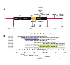
The binding of this antibody is dependent on the amino acids 333-357 of human NF-L, a region of a-helical coiled coil structure including a “stutter”, a region where the typical hydrophobic heptad repeat of coiled coils breaks down resulting in a slight kink in the structure. This antibody likely binds to a discontinuous epitope on the outside of the coiled coil dimer. This epitope is 100% conserved in all mammalian NF-L sequences.
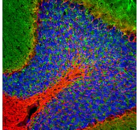
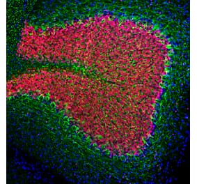
Immunofluorescent analysis of cow cerebellum section stained with Anti-NF-L Antibody [1B11] (A104310), at a dilution of 1:2,000, in green, and co-stained with Anti-Visinin Like Protein 1 Antibody (A85344), at a dilution of 1:2,000, in red. Blue is Hoechst staining of nuclear DNA. Small section of cow cerebellum was fixed in 4% paraformaldehyde for 3 days, cut to 45µM, and free-floating sections were stained with the above antibodies. Anti-NF-L Antibody [1B11] (A104310) labels dendrites and axons of neuronal cells in the granular layer (lower left) and prominent basket cell axons surrounding the large Purkinje neurons. Anti-Visinin Like Protein 1 Antibody (A85344) reveals protein expressed in granule cells and in synapses of the molecular layer of the cerebellum.
Western Blot - Anti-NF-L Antibody [1B11] (A104310)
Western blot analysis of different tissue lysates using Anti-NF-L Antibody [1B11] (A104310), at a dilution of 1:2,000, in green. The lanes contain: [Lane 1] protein standard, [Lane 2] rat brain , [Lane 3] mouse brain, and [Lane 4] cow cerebelum. Strong band at about 68-70kDa corresponds to NF-L protein; with the cow protein appearing slightly larger in molecular size as expected. Low molecular weight bands detected in cow brain sample are likely post mortem proteolytic forms of NF-L.
This antibody was made against NF-L purified from pig spinal cord and binds to amino acids 316-370 of human NF-L. The Anti-NF-L Antibody [1B11] (A104310) antibody has an epitope similar to the Uman NF-Light™ assay UD1, also known as 47.3. A shows diagrammatically the location of various NF-L antibody epitopes and B shows peptide sequence of the epitopes for our new antibodies.
Immunofluorescent analysis of cow cerebellum section stained with Anti-NF-L Antibody [1B11] (A104310) at a dilution of 1:2,000 (green), and costained with Anti-Visinin Like Protein 1 Antibody (A85344) at a dilution of 1:2,000 (red). Nuclei were stained with Hoechst (blue). Small section of cow cerebellum was fixed in 4% paraformaldehyde for 3 days, cut to 45µM, and free-floating sections were stained with the above antibodies. At this concentration the Anti-NF-L Antibody [1B11] (A104310) antibody labels dendrites and axons of neuronal cells in the granular layer (lower left) and prominent basket cell axons surrounding the large Purkinje neurons. The VLP1 antibody reveals protein expressed in granule cells and in synapses of the molecular layer of the cerebellum.
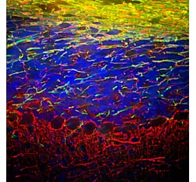
Immunostaining of a coronal section of the spinal cord of a rat given a midline C4 contusion injury three days previously. Sections were stained with Anti-NF-L Antibody (A270580) (red) and Anti-NF-L Antibody [1B11] (A104310) (green). Anti-NF-L Antibody [1B11] (A104310) stains prominent aggregates of material concentrated in the lateral funiculi and the dorsal columns but seen in lesser amounts throughout the section. These are degenerating and degenerated axons damaged by the C4 lesion. The Anti-NF-L Antibody antibody binds the C-terminal “tail” region of NF-L which is absent or destroyed during degeneration, so the positive profiles are largely negative for Anti-NF-L Antibody.
Immunohistochemistry analysis of a formalin fixed paraffin embedded brain stem section from a transgenic mouse model of ALS stained with Anti-NF-L Antibody [1B11] (A104310) at a dilution of 1:1,000 detected in DAB (brown) following the Vector Labs Mouse on Mouse ImmPRESS® HRP method, without the antigen retrieval step. Counterstained with Hematoxylin (blue). The Anti-NF-L Antibody [1B11] (A104310) labels what are clearly degenerated axons, showing typical swollen, sinusoidal and discontinuous profiles. Note that under these conditions healthy axons are not stained.
Western Blot - Anti-NF-L Antibody [1B11] (A104310)
Western blots of Uman NF-LIGHT™ antibodies and a set of reagents on Recombinant Human NF-L Protein (A270573) and Recombinant Human NF L Protein (A333288). The lanes contain: [Lane 1] protein standards (red), [Lane 2] Recombinant Human NF-L Protein (A270573), [Lane 3] Recombinant Human NF L Protein (A333288). Recombinant Human NF-L Protein (A270573) runs at about 75kDa, while Recombinant Human NF L Protein (A333288) runs at about 12kDa. All five antibodies recognize both constructs. UD1 is also known as 2.1 is the detection reagent in the Uman NF-LIGHT™ assay while UD2, also known as 47.3 is the capture reagent (6). The three other lanes show results obtained with Anti-NF-L Antibody [1B11] (A104310) and Anti-NF-L Antibody [6H63] (A333276) respectively as indicated. All these antibodies binds to an epitope flanking the so-called second “stutter” in the Coil 2 region of the a-helical coiled coil “rod” region of NF-L. The binding properties are very similar to the Uman mouse monoclonal antibody UD1 also known as 2.1, as described originally in Norgren et al. 2002. This antibody is the key capture reagent in the NF-Light™ assay of Uman Diagnostics and the Quanterix Simoa™ NF-L assay.


![Immunofluorescence - Anti-NF-L Antibody [1B11] (A104310) - Antibodies.com](https://cdn.antibodies.com/image/catalog/104/A104310_1.jpg?profile=product_top)
![Western Blot - Anti-NF-L Antibody [1B11] (A104310) - Antibodies.com](https://cdn.antibodies.com/image/catalog/104/A104310_2.jpg?profile=product_top)
![Epitope Diagram - Anti-NF-L Antibody [1B11] (A104310) - Antibodies.com](https://cdn.antibodies.com/image/catalog/104/A104310_3.jpg?profile=product_top)
![Immunofluorescence - Anti-NF-L Antibody [1B11] (A104310) - Antibodies.com](https://cdn.antibodies.com/image/catalog/104/A104310_4.jpg?profile=product_top)
![Immunofluorescence - Anti-NF-L Antibody [1B11] (A104310) - Antibodies.com](https://cdn.antibodies.com/image/catalog/104/A104310_5.jpg?profile=product_top)
![Immunohistochemistry - Anti-NF-L Antibody [1B11] (A104310) - Antibodies.com](https://cdn.antibodies.com/image/catalog/104/A104310_6.jpg?profile=product_top)
![Western Blot - Anti-NF-L Antibody [1B11] (A104310) - Antibodies.com](https://cdn.antibodies.com/image/catalog/104/A104310_7.jpg?profile=product_top)
![Immunofluorescence - Anti-NF-L Antibody [1B11] (A104310) - Antibodies.com](https://cdn.antibodies.com/image/catalog/104/A104310_1.jpg?profile=product_top_thumb)
![Western Blot - Anti-NF-L Antibody [1B11] (A104310) - Antibodies.com](https://cdn.antibodies.com/image/catalog/104/A104310_2.jpg?profile=product_top_thumb)
![Epitope Diagram - Anti-NF-L Antibody [1B11] (A104310) - Antibodies.com](https://cdn.antibodies.com/image/catalog/104/A104310_3.jpg?profile=product_top_thumb)
![Immunofluorescence - Anti-NF-L Antibody [1B11] (A104310) - Antibodies.com](https://cdn.antibodies.com/image/catalog/104/A104310_4.jpg?profile=product_top_thumb)
![Immunofluorescence - Anti-NF-L Antibody [1B11] (A104310) - Antibodies.com](https://cdn.antibodies.com/image/catalog/104/A104310_5.jpg?profile=product_top_thumb)
![Immunohistochemistry - Anti-NF-L Antibody [1B11] (A104310) - Antibodies.com](https://cdn.antibodies.com/image/catalog/104/A104310_6.jpg?profile=product_top_thumb)
![Western Blot - Anti-NF-L Antibody [1B11] (A104310) - Antibodies.com](https://cdn.antibodies.com/image/catalog/104/A104310_7.jpg?profile=product_top_thumb)
![Immunofluorescence - Anti-NF-L Antibody [1B11] (A104310) - Antibodies.com](https://cdn.antibodies.com/image/catalog/104/A104310_1.jpg?profile=product_image)
![Western Blot - Anti-NF-L Antibody [1B11] (A104310) - Antibodies.com](https://cdn.antibodies.com/image/catalog/104/A104310_2.jpg?profile=product_image)
![Epitope Diagram - Anti-NF-L Antibody [1B11] (A104310) - Antibodies.com](https://cdn.antibodies.com/image/catalog/104/A104310_3.jpg?profile=product_image)
![Immunofluorescence - Anti-NF-L Antibody [1B11] (A104310) - Antibodies.com](https://cdn.antibodies.com/image/catalog/104/A104310_4.jpg?profile=product_image)
![Immunofluorescence - Anti-NF-L Antibody [1B11] (A104310) - Antibodies.com](https://cdn.antibodies.com/image/catalog/104/A104310_5.jpg?profile=product_image)
![Immunohistochemistry - Anti-NF-L Antibody [1B11] (A104310) - Antibodies.com](https://cdn.antibodies.com/image/catalog/104/A104310_6.jpg?profile=product_image)
![Western Blot - Anti-NF-L Antibody [1B11] (A104310) - Antibodies.com](https://cdn.antibodies.com/image/catalog/104/A104310_7.jpg?profile=product_image)
![Immunofluorescence - Anti-NF-L Antibody [DA2] (A85454) - Antibodies.com](https://cdn.antibodies.com/image/catalog/85/A85454_1.jpg?profile=product_alternative)

![Immunofluorescence - Anti-NF-L Antibody [7D1] (A85453) - Antibodies.com](https://cdn.antibodies.com/image/catalog/85/A85453_2.jpg?profile=product_alternative)
![Immunohistochemistry - Anti-NF-L Antibody [NR-4] (A252677) - Antibodies.com](https://cdn.antibodies.com/image/catalog/249/A249494_1.jpg?profile=product_alternative)
![Immunohistochemistry - Anti-NF-L Antibody [NR-4] - BSA and Azide free (A249494) - Antibodies.com](https://cdn.antibodies.com/image/catalog/252/A252674_1.jpg?profile=product_alternative)

![Immunohistochemistry - Anti-NF-L Antibody [NFL/736] - BSA and Azide free (A249496) - Antibodies.com](https://cdn.antibodies.com/image/catalog/252/A252676_1.jpg?profile=product_alternative)
![Epitope Diagram - Anti-NF-L Antibody [6H112] (A270560) - Antibodies.com](https://cdn.antibodies.com/image/catalog/270/A270560_1.jpg?profile=product_alternative)