Immunofluorescent analysis of mouse cerebellum section stained with Anti-NF-L Antibody, at a dilution of 1:5,000, in red, and Anti-MBP Antibody (A85321 | 1:5,000, in green. Following transcardial perfusion of mouse with 4% paraformaldehyde, brain was post fixed for 24 hours, cut to 45µM, and free-floating sections were stained with the above antibodies. Anti-NF-L Antibody labels dendrites and axons of neuronal cells, and Anti-MBP Antibody stains the network of myelin sheathes around axons.
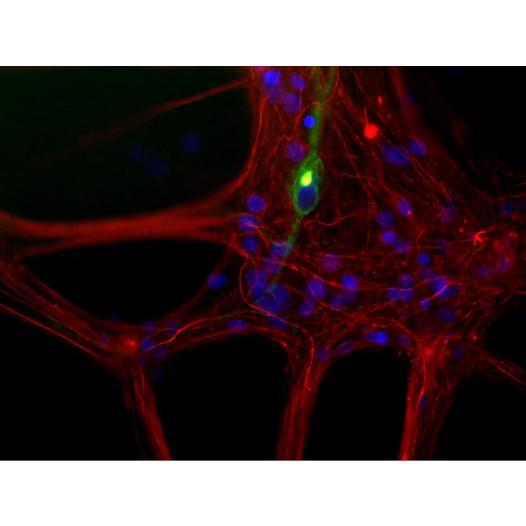
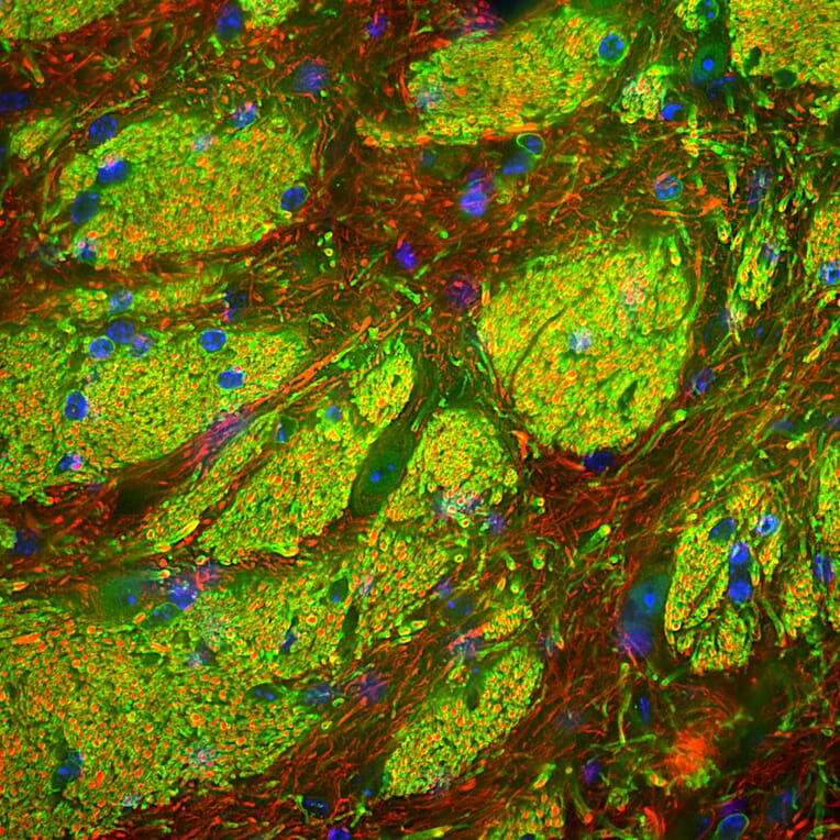
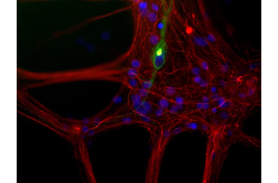
Mixed neuron/glia cultures from newborn rat brain stained with Anti-Peripherin Antibody (A85432 | green) and Anti-NF-L Antibody (red). A class of large neurons, like the one in the middle of this image, contains peripherin, while the majority of neurons and their processes contain NF-L and not peripherin. Interestingly, the peripherin positive cells often contain a cytoplasmic inclusion next to the nucleus which stains for both peripherin and NF-L, and so appears golden in this image. The blue channel reveals the localization of DNA.
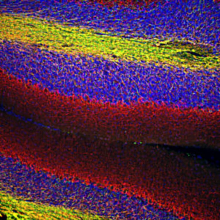
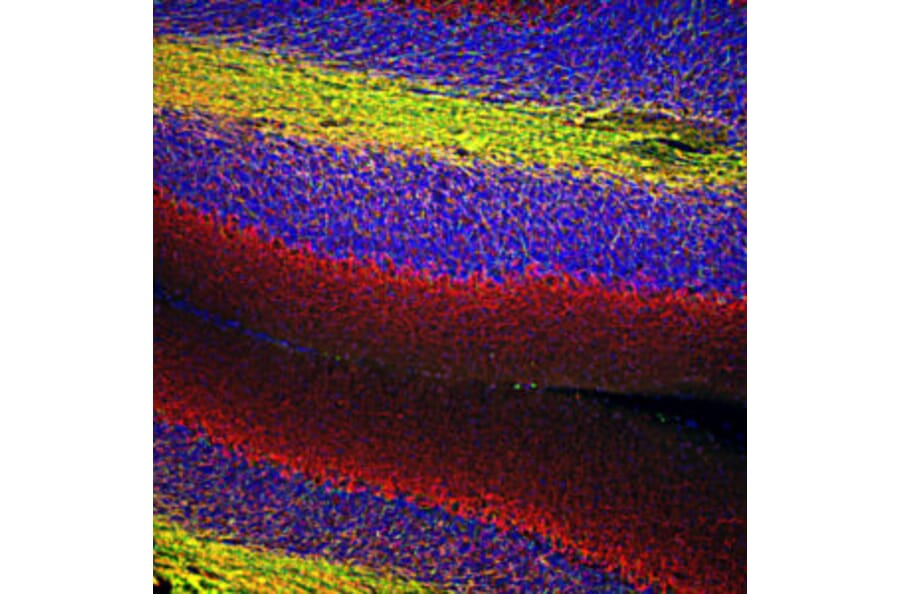
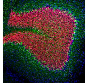
Immunofluorescent analysis of rat brain cerebellum section stained with Anti-Myelin Basic Protein Antibody (A85321) at a dilution of 1:5,000 (green) and costained with Anti-NF-L Antibody (A85451) at a dilution of 1:5,000 (red). This antibody stains oligodendrocytes and the myelin sheathes around axons. NF-L antibody labels dendrites and axons of neuronal cells in the molecular and granule layers.
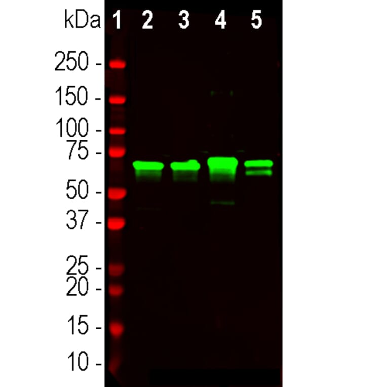
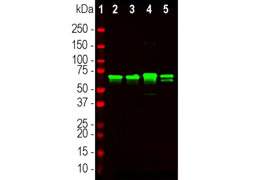
Western blot analysis of different tissue lysates using Anti-NF-L Antibody (A85451), at a dilution of 1:20,000, in green. The lanes contain samples from: [Lane 1] Protein standards, in red, [Lane 2] rat brain, [Lane 3] rat spinal cord, [Lane 4] mouse brain, and [Lane 5] mouse spinal cord. The strong band at 68 kDa corresponds to the NF-L protein.
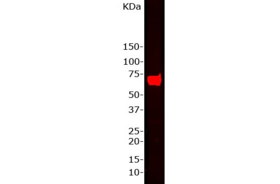
Western blot of whole rat brain homogenate stained with Anti-NF-L Antibody, at a dilution of 1:15,000). A prominent band running with an apparent SDS-PAGE molecular weight of ~68 kDa corresponds to rodent NF-L.
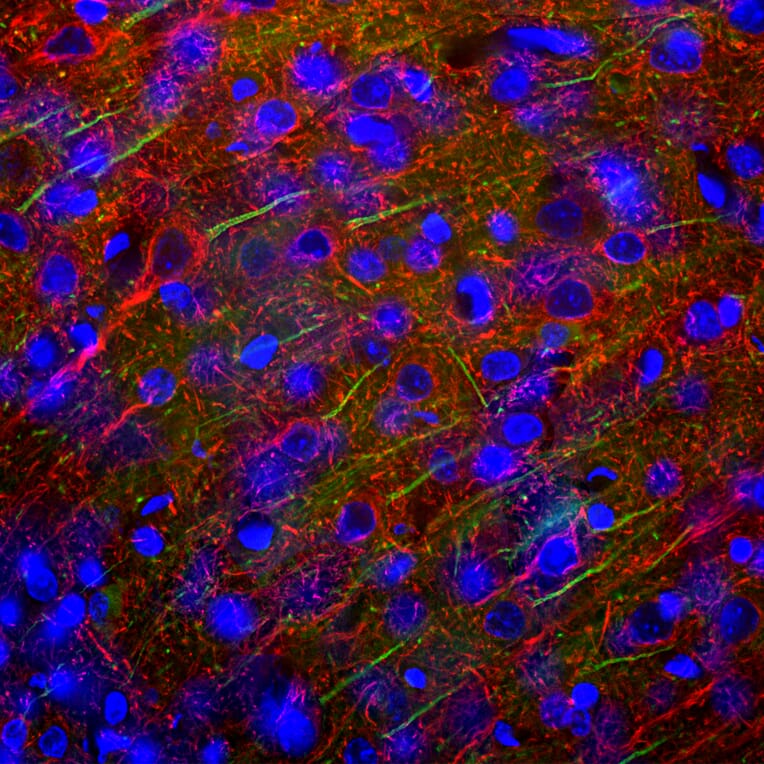
Immunofluorescent analysis of rat cerebellum section stained with Anti-NF-L Antibody (A85451), at a dilution of 1:5,000, in red, and co-stained with Anti-beta Synuclein Antibody [6A10] (A270558), at a dilution of 1:500, in green. The nuclear DNA is visualised in blue using Hoechst staining. Following transcardial perfusion of the rat with 4% paraformaldehyde, the brain was post-fixed for 24 hours, cut to 45 µm, and free-floating sections were stained with the above antibodies. The Anti-NF-L Antibody (A85451) labels dendrites and axons of neuronal cells, and the Anti-beta Synuclein Antibody [6A10] (A270558) detects protein that is concentrated in synaptic regions.
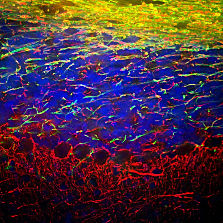
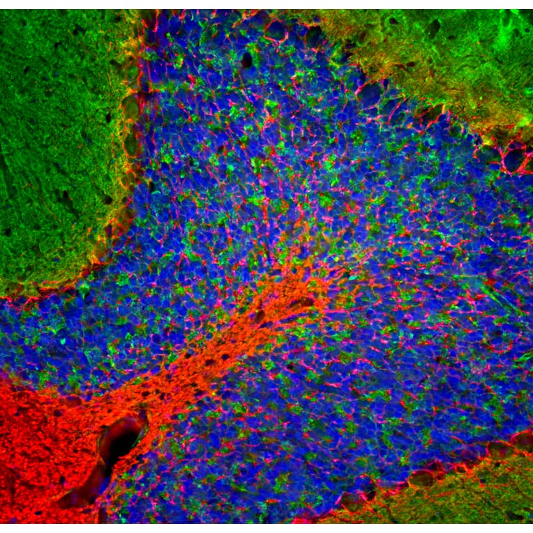
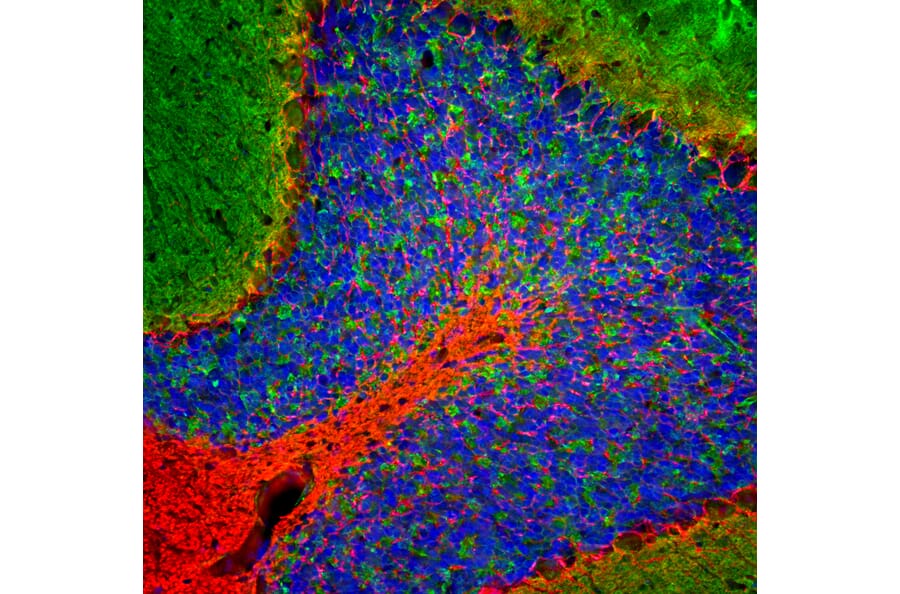
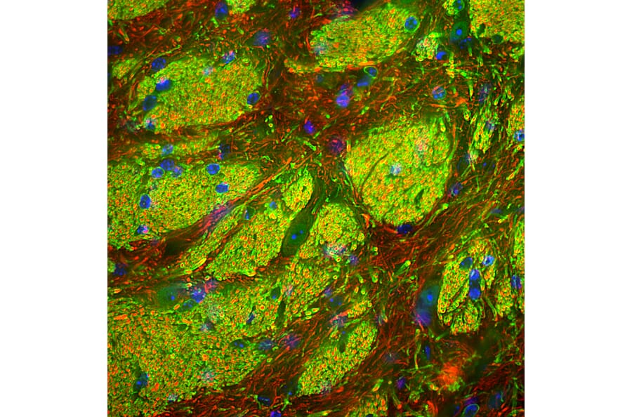
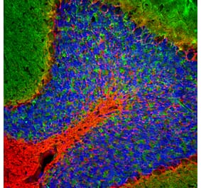
Immunofluorescent analysis of mouse striatum section stained using Anti-CNPase Antibody (A270543), at a dilution of 1:3,000, in green, and co-stained with Anti-NF-L Antibody (A85451), at a dilution of 1:5,000, in red. Nuclear DNA is visualised in blue using Hoechst staining. Following transcardial perfusion of the mouse with 4% paraformaldehyde, the brain was post-fixed for 24 hours, cut to 45 µm, and free-floating sections were stained with the above antibodies. Anti-CNPase Antibody (A270543) stains myelin sheath and the plasma membranes of oligodendrocytes; the myelin producing cells of the CNS. Anti-NF-L Antibody (A85451) labels axons of neuronal cells enclosed by the myelin.
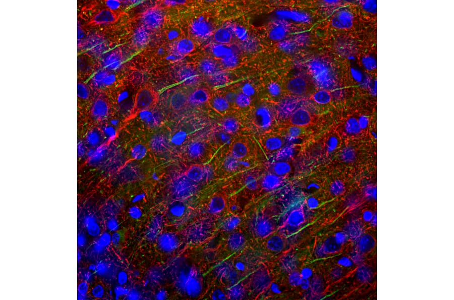
Immunofluorescent analysis of rat cortex section stained with Anti-Ankyrin 3 Antibody [2A8], at a dilution of 1:2,000, in green, and co-stained with Anti-NF-L Antibody (A85451), at a dilution of 1:5,000, in red. Nuclear DNA is visualised in blue using Hoechst staining. Following transcardial perfusion with 4% paraformaldehyde, the brain was post-fixed for 24 hours, cut to 45 µm, and free-floating sections were stained using the above antibodies. Anti-Ankyrin 3 Antibody [2A8] stains the axonal initial segments, while Anti-NF-L Antibody (A85451) labels dendrites and axons of neuronal cells.
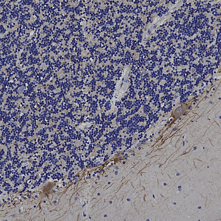
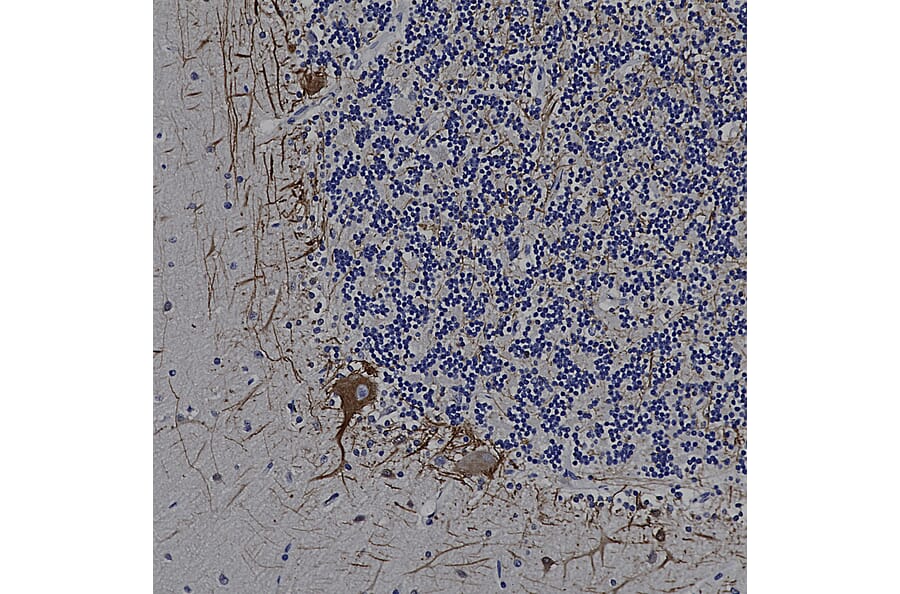
Immunohistochemistry analysis of a formalin fixed paraffin embedded human cerebellum section with Anti-NF-L Antibody (A85451) at a dilution of 1:5,000. Anti-NF-L Antibody (A85451) labels the perinkarya and dendrites of Purkinje cells and the projections of neuronal cells within the granular layer. Note: this antibody performs well in testing with both 4% PFA and standard NBF fixed rat, mouse and human tissues.
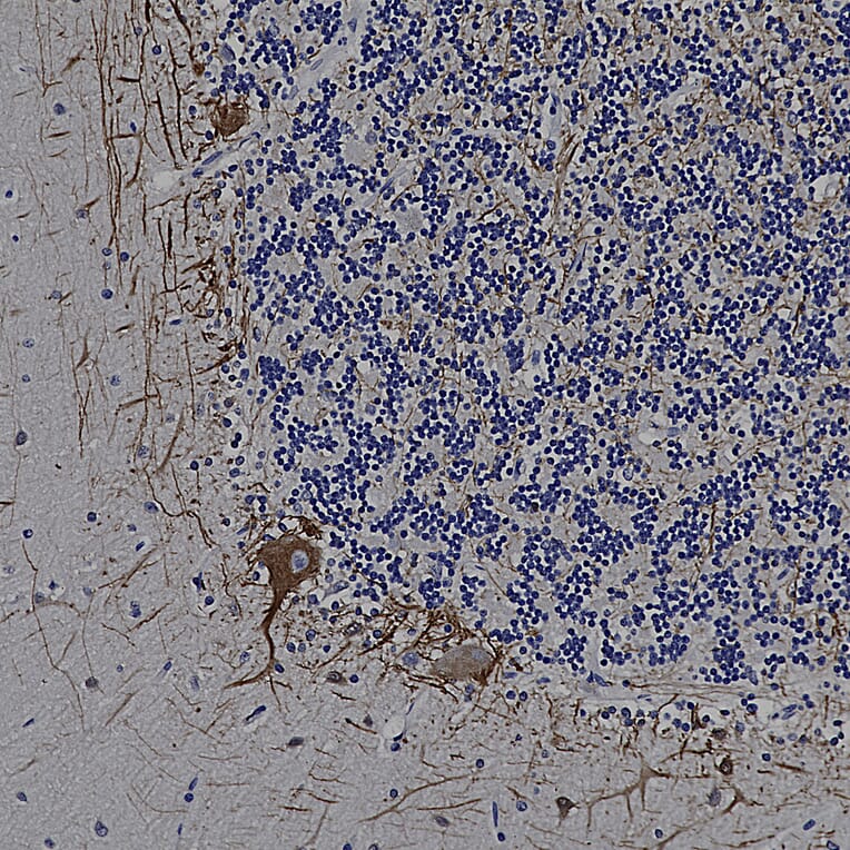
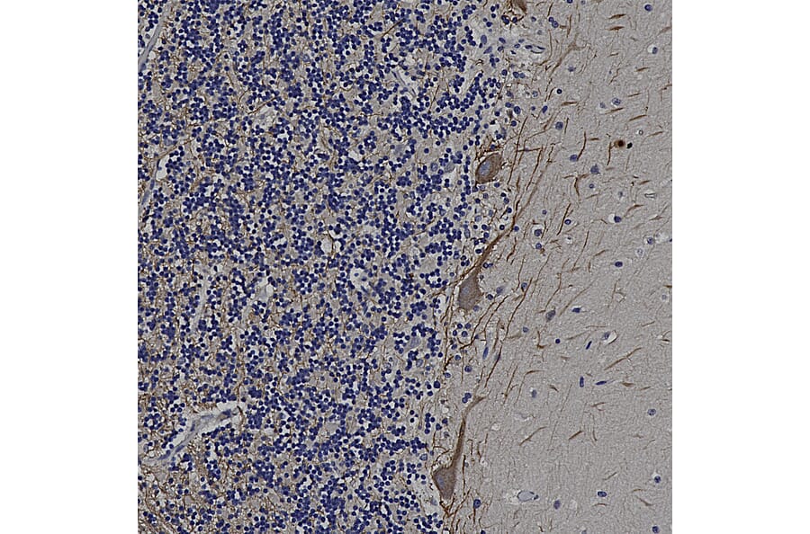
Immunohistochemistry analysis of a formalin fixed paraffin embedded human cerebellum section with Anti-NF-M Antibody (A85323) at a dilution of 1:1,000 detected with DAB (brown) using the Vector Labs ImmPRESS method and reagents with citra buffer retrieval. Counterstained with Hematoxylin (blue). Anti-NF-L Antibody (A85451) labels the perinkarya and dendrites of Purkinje cells and the projections of neuronal cells within the granular layer. Note: this antibody performs well in testing with both 4% PFA and standard NBF fixed rat, mouse, and human tissues.
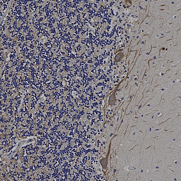
Immunohistochemistry analysis of a formalin fixed paraffin embedded human cerebellum section with Anti-NF-M Antibody (A85323) at a dilution of 1:2,000 detected with DAB (brown) using the Vector Labs ImmPRESS method and reagents with citra buffer retrieval. Counterstained with Hematoxylin (blue). Anti-NF-L Antibody (A85451) labels the perinkarya and dendrites of Purkinje cells and the projections of neuronal cells within the granular layer. Note: this antibody performs well in testing with both 4% PFA and standard NBF fixed rat, mouse, and human tissues.


![Immunofluorescence - Anti-NF-L Antibody [DA2] (A85454) - Antibodies.com](https://cdn.antibodies.com/image/catalog/85/A85454_1.jpg?profile=product_alternative)

![Immunofluorescence - Anti-NF-L Antibody [1B11] (A104310) - Antibodies.com](https://cdn.antibodies.com/image/catalog/104/A104310_1.jpg?profile=product_alternative)
![Immunofluorescence - Anti-NF-L Antibody [7D1] (A85453) - Antibodies.com](https://cdn.antibodies.com/image/catalog/85/A85453_2.jpg?profile=product_alternative)
![Immunohistochemistry - Anti-NF-L Antibody [NR-4] (A252677) - Antibodies.com](https://cdn.antibodies.com/image/catalog/249/A249494_1.jpg?profile=product_alternative)
![Immunohistochemistry - Anti-NF-L Antibody [NR-4] - BSA and Azide free (A249494) - Antibodies.com](https://cdn.antibodies.com/image/catalog/252/A252674_1.jpg?profile=product_alternative)

![Immunohistochemistry - Anti-NF-L Antibody [NFL/736] - BSA and Azide free (A249496) - Antibodies.com](https://cdn.antibodies.com/image/catalog/252/A252676_1.jpg?profile=product_alternative)
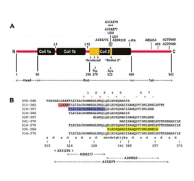
![Epitope Diagram - Anti-NF-L Antibody [6H112] (A270560) - Antibodies.com](https://cdn.antibodies.com/image/catalog/270/A270560_1.jpg?profile=product_alternative)