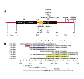
The epitope for this antibody is mapped to a short peptide in the C-terminal “tail” region of the molecule, within the sequence SYYTSHVQEEQIEVE, amino acids 441-460 of human NF-L.
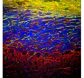
Immunohistochemical analysis of a neuron in cell culture stained with Anti-Neurofilament Light Polypeptide Antibody [DA2] (A85454), in green. The culture was derived from adult rat cortex, grown under conditions to induce neuronal survival and differentiation. The culture was counterstained with Anti-alpha Internexin Antibody (A85441), in red; this antibody highlights a network of small neurons at early stages of development.
Western blot analysis of whole tissue lysates using Anti-Neurofilament Light Polypeptide Antibody [DA2] (A85454), dilution 1:5,000, in green. The lanes contain: [Lane 1] protein standard (red), [Lane 2] rat brain, [Lane 3] rat spinal cord, [Lane 4] mouse brain, [Lane 5] mouse spinal cord. The strong band at 68-70 kDa corresponds to the NF-L protein.
Rat spinal cord homogenate showing the major intermediate filament proteins of the nervous system (Lane 1). The remaining lanes show blots of this material stained with various antibodies including Anti-NF-L Antibody (Lane 4).
Immunofluorescent analysis of rat frontal cortex section stained with Anti-NF-L Antibody [DA2] (A85454) at a dilution of 1:500 (red) and costained with Anti-GFAP Antibody (A85307) at a dilution of 1:5,000 (green). Following transcardial perfusion of rat with 4% paraformaldehyde, brain was post fixed for 24 hours, cut to 45µM, and free-floating sections were stained with above antibodies. This antibody labels cell bodies and processes of pyramidal neurons, as well as dendrites and axons of other neuronal cells, while the GFAP antibody stains the network of glial cells.
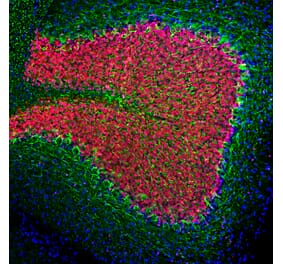
Immunohistochemistry analysis of a formalin fixed paraffin embedded human cerebellum section with Anti-NF-L Antibody [DA2] (A85454) at a dilution of 1:2,000 detected in DAB (brown) following the ImmPress method with citra buffer retrieval. Counterstained with Hematoxylin (blue). Anti-NF-L Antibody [DA2] (A85454) labels neuronal cells and their processes. Note: this antibody performs well in testing with both 4% PFA and standard NBF fixed tissues and is the recommended clone for use in NF-L immunostaining of rat tissue. Mouse and human tissue has also been validated in paraffin immuno-histochemistry with this antibody.
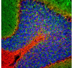
Immunohistochemistry analysis of a formalin fixed paraffin embedded human cerebellum section with Anti-NF-L Antibody [DA2] (A85454) at a dilution of 1:2,000 detected with DAB (brown) using the Vector Labs ImmPRESS method and reagents with Borg retrieval. Counterstained with Hematoxylin (blue). The Anti-NF-L Antibody [DA2] (A85454) strongly labels the perikarya and processes of Purkinje cells. Note: this antibody performs well in testing with both 4% PFA and NBF fixed rat tissues but requires high pH retrieval to stain long term NBF fixed human tissue effectively.


![Immunofluorescence - Anti-NF-L Antibody [DA2] (A85454) - Antibodies.com](https://cdn.antibodies.com/image/catalog/85/A85454_1.jpg?profile=product_top)
![Western Blot - Anti-NF-L Antibody [DA2] (A85454) - Antibodies.com](https://cdn.antibodies.com/image/catalog/85/A85454_2.jpg?profile=product_top)
![Epitope Diagram - Anti-NF-L Antibody [DA2] (A85454) - Antibodies.com](https://cdn.antibodies.com/image/catalog/85/A85454_3.jpg?profile=product_top)
![Western Blot - Anti-NF-L Antibody [DA2] (A85454) - Antibodies.com](https://cdn.antibodies.com/image/catalog/85/A85454_4.jpg?profile=product_top)
![Immunofluorescence - Anti-NF-L Antibody [DA2] (A85454) - Antibodies.com](https://cdn.antibodies.com/image/catalog/85/A85454_5.jpg?profile=product_top)
![Immunohistochemistry - Anti-NF-L Antibody [DA2] (A85454) - Antibodies.com](https://cdn.antibodies.com/image/catalog/85/A85454_6.jpg?profile=product_top)
![Immunohistochemistry - Anti-NF-L Antibody [DA2] (A85454) - Antibodies.com](https://cdn.antibodies.com/image/catalog/85/A85454_7.jpg?profile=product_top)
![Immunofluorescence - Anti-NF-L Antibody [DA2] (A85454) - Antibodies.com](https://cdn.antibodies.com/image/catalog/85/A85454_1.jpg?profile=product_top_thumb)
![Western Blot - Anti-NF-L Antibody [DA2] (A85454) - Antibodies.com](https://cdn.antibodies.com/image/catalog/85/A85454_2.jpg?profile=product_top_thumb)
![Epitope Diagram - Anti-NF-L Antibody [DA2] (A85454) - Antibodies.com](https://cdn.antibodies.com/image/catalog/85/A85454_3.jpg?profile=product_top_thumb)
![Western Blot - Anti-NF-L Antibody [DA2] (A85454) - Antibodies.com](https://cdn.antibodies.com/image/catalog/85/A85454_4.jpg?profile=product_top_thumb)
![Immunofluorescence - Anti-NF-L Antibody [DA2] (A85454) - Antibodies.com](https://cdn.antibodies.com/image/catalog/85/A85454_5.jpg?profile=product_top_thumb)
![Immunohistochemistry - Anti-NF-L Antibody [DA2] (A85454) - Antibodies.com](https://cdn.antibodies.com/image/catalog/85/A85454_6.jpg?profile=product_top_thumb)
![Immunohistochemistry - Anti-NF-L Antibody [DA2] (A85454) - Antibodies.com](https://cdn.antibodies.com/image/catalog/85/A85454_7.jpg?profile=product_top_thumb)
![Immunofluorescence - Anti-NF-L Antibody [DA2] (A85454) - Antibodies.com](https://cdn.antibodies.com/image/catalog/85/A85454_1.jpg?profile=product_image)
![Western Blot - Anti-NF-L Antibody [DA2] (A85454) - Antibodies.com](https://cdn.antibodies.com/image/catalog/85/A85454_2.jpg?profile=product_image)
![Epitope Diagram - Anti-NF-L Antibody [DA2] (A85454) - Antibodies.com](https://cdn.antibodies.com/image/catalog/85/A85454_3.jpg?profile=product_image)
![Western Blot - Anti-NF-L Antibody [DA2] (A85454) - Antibodies.com](https://cdn.antibodies.com/image/catalog/85/A85454_4.jpg?profile=product_image)
![Immunofluorescence - Anti-NF-L Antibody [DA2] (A85454) - Antibodies.com](https://cdn.antibodies.com/image/catalog/85/A85454_5.jpg?profile=product_image)
![Immunohistochemistry - Anti-NF-L Antibody [DA2] (A85454) - Antibodies.com](https://cdn.antibodies.com/image/catalog/85/A85454_6.jpg?profile=product_image)
![Immunohistochemistry - Anti-NF-L Antibody [DA2] (A85454) - Antibodies.com](https://cdn.antibodies.com/image/catalog/85/A85454_7.jpg?profile=product_image)
![Immunofluorescence - Anti-NF-L Antibody [1B11] (A104310) - Antibodies.com](https://cdn.antibodies.com/image/catalog/104/A104310_1.jpg?profile=product_alternative)

![Immunofluorescence - Anti-NF-L Antibody [7D1] (A85453) - Antibodies.com](https://cdn.antibodies.com/image/catalog/85/A85453_2.jpg?profile=product_alternative)
![Immunohistochemistry - Anti-NF-L Antibody [NR-4] - BSA and Azide free (A249494) - Antibodies.com](https://cdn.antibodies.com/image/catalog/252/A252674_1.jpg?profile=product_alternative)

![Immunohistochemistry - Anti-NF-L Antibody [NR-4] (A252677) - Antibodies.com](https://cdn.antibodies.com/image/catalog/249/A249494_1.jpg?profile=product_alternative)
![Immunohistochemistry - Anti-NF-L Antibody [NFL/736] - BSA and Azide free (A249496) - Antibodies.com](https://cdn.antibodies.com/image/catalog/252/A252676_1.jpg?profile=product_alternative)
![Epitope Diagram - Anti-NF-L Antibody [6H112] (A270560) - Antibodies.com](https://cdn.antibodies.com/image/catalog/270/A270560_1.jpg?profile=product_alternative)