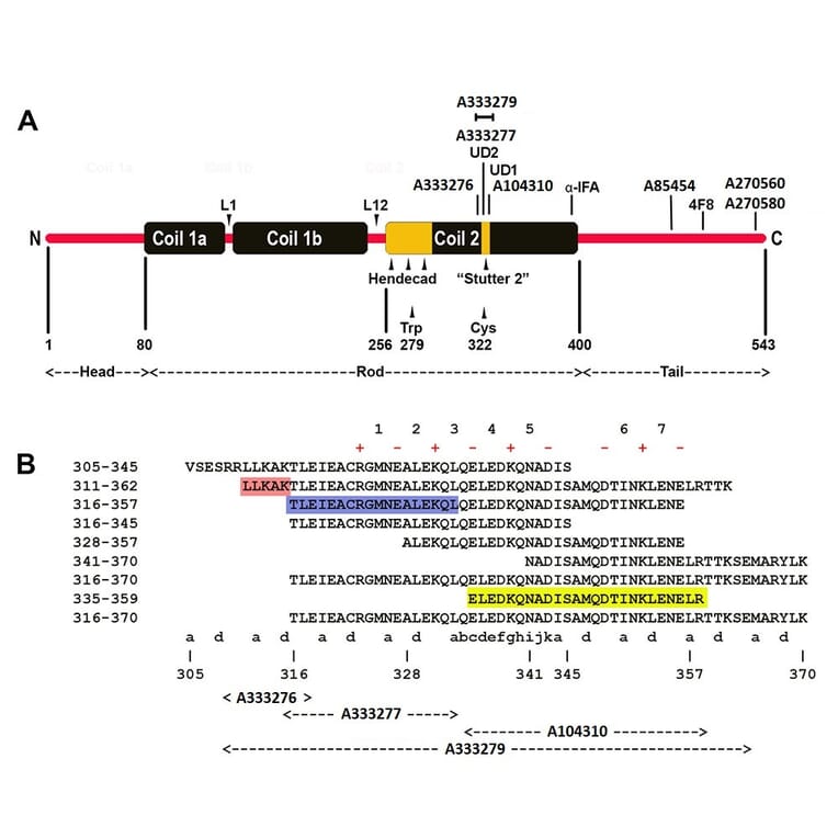
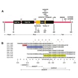
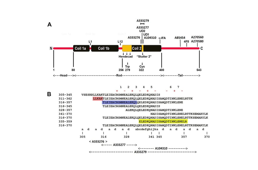
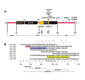
Epitope map of novel DegenoTag™antibodies on human NF-L including Anti-NF-L Antibody [6H63] (A333276). We mapped the epitopes for the two mouse monoclonal antibodies to NF-L used in the Uman Diagnostics NF-LIGHT™ assay. In the diagram these are indicated as “UD1” and UD2”. UD1 is also known as 2.1 and serves as the NF-L detection reagent while UD2 is also known as 47.3 and is the capture reagent. The same pair of antibodies are used in the Simoa™ NF-L assay of Quanterix. These are both located in a short peptide flanking the “stutter 2” region of the a-helical “Coil 2” region. Our own antibodies were made against NF-L 311-362 and share the degeneration specific staining patter shown by UD1 and UD2. The epitope for Anti-NF-L Antibody [6H63] (A333276) is heavily dependent on the peptide LLKAK highlighted on the figure.
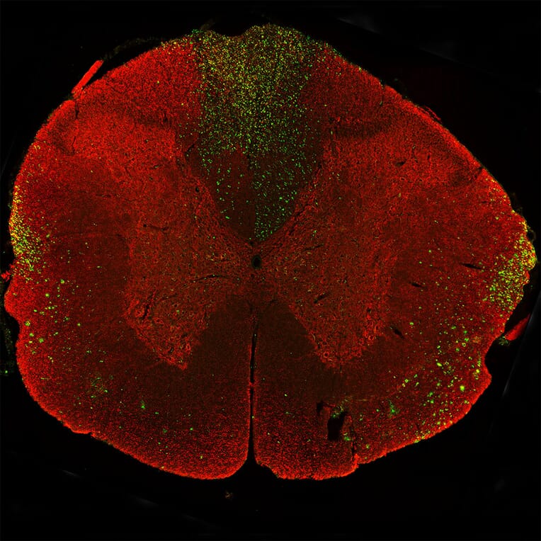
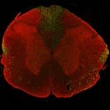
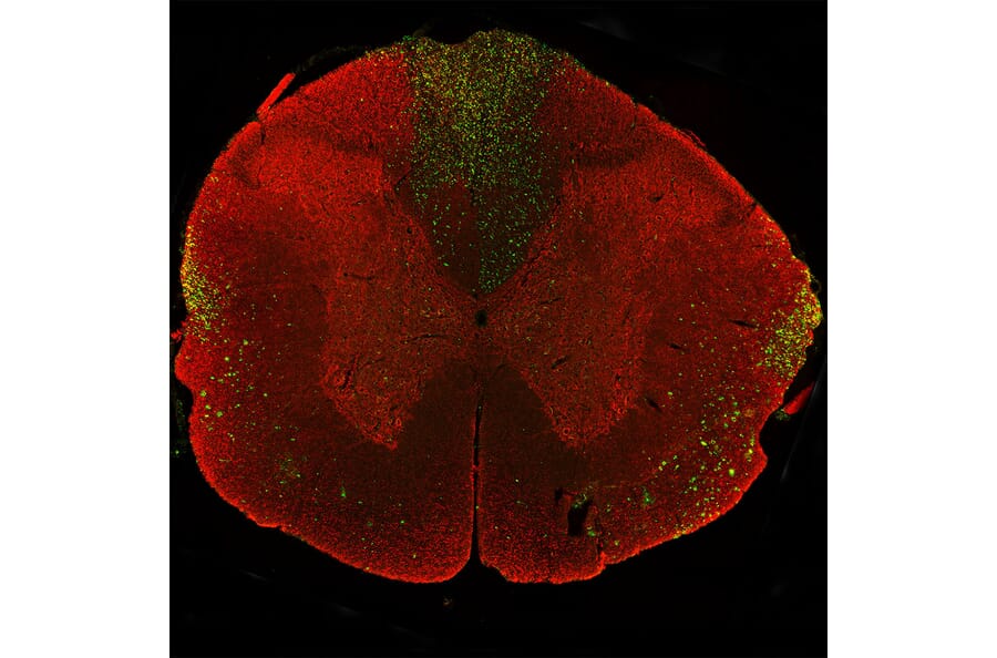
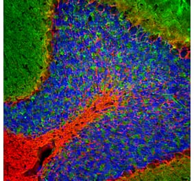
Immunostaining of a coronal section of the spinal cord of a rat given a midline C4 contusion injury three days previously. Sections were stained with Anti-NF-L Antibody (A270580) (red) and Anti-NF-L Antibody [6H63] (A333276) (green). Anti-NF-L Antibody [6H63] (A333276) stains prominent aggregates of material concentrated in the lateral funiculi and the dorsal columns but seen in lesser amounts throughout the section. These are degenerating and degenerated axons damaged by the C4 lesion. The Anti-NF-L Antibody antibody binds the C-terminal “tail” region of NF-L which is absent or destroyed during degeneration, so the positive profiles are largely negative for Anti-NF-L Antibody.
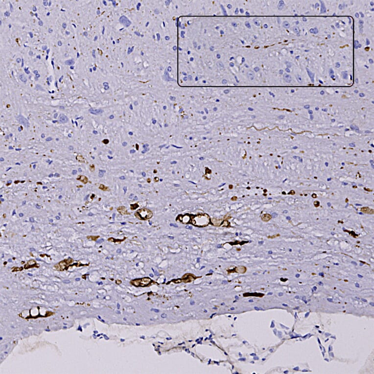
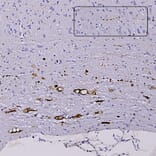
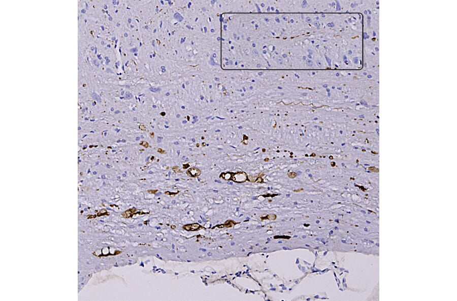
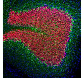
Immunohistochemistry analysis of formalin fixed paraffin embedded sections of heterozygous G85R-SOD1-YFP mice injected with mutant SOD1 in the hind limb using Anti-NF-L Antibody [6H63] (A333276) and degenerated processes were seen in the sciatic nerve, in fibers in the spinal cord and as shown here in the brain stem. Note swollen and sinusoidal profiles. The inset shows an example of a beaded profile typical of degenerating processes. Staining was performed using Vector Labs mouse on mouse Immpress method and reagents following protocol.
Western Blot - Anti-NF-L Antibody [6H63] (A333276)
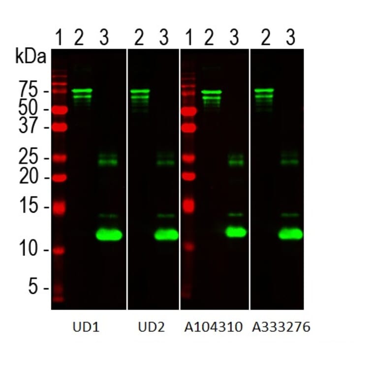
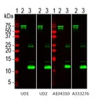
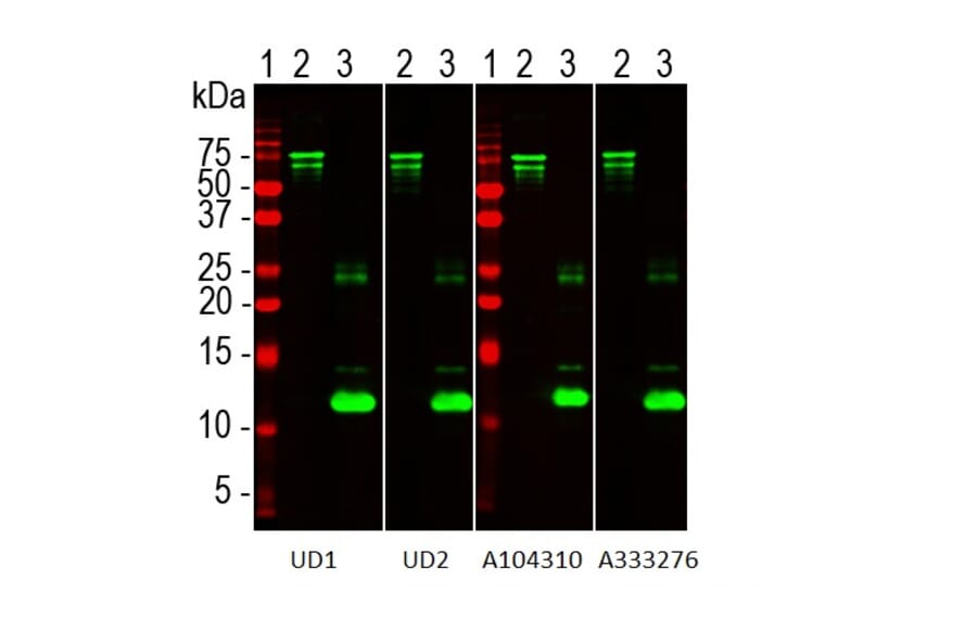
Western blots of Uman NF-LIGHT™ antibodies and a set of reagents on Recombinant Human NF-L Protein (A270573) and Recombinant Human NF L Protein (A333288). The lanes contain: [Lane 1] protein standard (red), [Lane 2] Recombinant Human NF-L Protein (A270573), [Lane 3] Recombinant Human NF L Protein (A333288). Recombinant Human NF-L Protein (A270573) runs at about 75kDa, while Recombinant Human NF L Protein (A333288) runs at about 12kDa. All five antibodies recognize both constructs. UD1 is also known as 2.1 is the detection reagent in the Uman NF-LIGHT™ assay while UD2, also known as 47.3 is the capture reagent (6). The three other lanes show results obtained with Anti-NF-L Antibody [1B11] (A104310) and Anti-NF-L Antibody [6H63] (A333276) respectively as indicated. All these antibodies binds to an epitope flanking the so-called second “stutter” in the Coil 2 region of the a-helical coiled coil “rod” region of NF-L. Anti-NF-L Antibody [6H63] (A333276) binds an epitope just N-terminal to the Uman antibody UD2.
Western Blot - Anti-NF-L Antibody [6H63] (A333276)
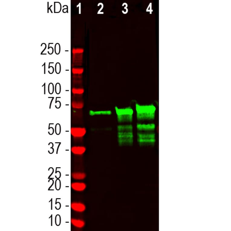
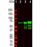
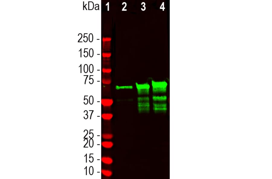
Western blot analysis of crude CNS homogenates using Anti-NF-L Antibody [6H63] (A333276). The lanes contain: [Lane 1] protein standards, [Lane 2] homogenate of E20 rat spinal cord, [Lane 3] homogenate of adult rat spinal cord, and [Lane 4] homogenate of cow spinal cord extract. Anti-NF-L Antibody [6H63] (A333276) binds the denatured forms of NF-L with apparent molecular weight 68-70kDa as immobilized on western blotting membranes. Lower molecular weight bands under the major band are proteolytic fragments of NF-L.


![Immunofluorescence - Anti-NF-L Antibody [DA2] (A85454) - Antibodies.com](https://cdn.antibodies.com/image/catalog/85/A85454_1.jpg?profile=product_alternative)

![Immunofluorescence - Anti-NF-L Antibody [1B11] (A104310) - Antibodies.com](https://cdn.antibodies.com/image/catalog/104/A104310_1.jpg?profile=product_alternative)
![Immunofluorescence - Anti-NF-L Antibody [7D1] (A85453) - Antibodies.com](https://cdn.antibodies.com/image/catalog/85/A85453_2.jpg?profile=product_alternative)
![Immunohistochemistry - Anti-NF-L Antibody [NR-4] (A252677) - Antibodies.com](https://cdn.antibodies.com/image/catalog/249/A249494_1.jpg?profile=product_alternative)
![Immunohistochemistry - Anti-NF-L Antibody [NR-4] - BSA and Azide free (A249494) - Antibodies.com](https://cdn.antibodies.com/image/catalog/252/A252674_1.jpg?profile=product_alternative)
![Immunohistochemistry - Anti-NF-L Antibody [NFL/736] - BSA and Azide free (A249496) - Antibodies.com](https://cdn.antibodies.com/image/catalog/252/A252676_1.jpg?profile=product_alternative)
![Epitope Diagram - Anti-NF-L Antibody [6H112] (A270560) - Antibodies.com](https://cdn.antibodies.com/image/catalog/270/A270560_1.jpg?profile=product_alternative)