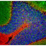
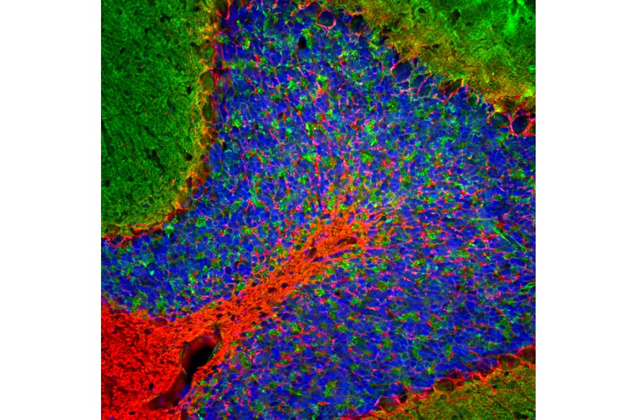
Immunofluorescent analysis of rat cerebellum section stained with Anti-NF-L Antibody (A270580), at a dilution of 1:5,000, in red, and co-stained with Anti-beta Synuclein Antibody [6A10] (A270558), at a dilution of 1:500, in green. Nuclear DNA is visualised in blue using Hoechst staining. Following transcardial perfusion with 4% paraformaldehyde, the brain was post-fixed for 24 hours, cut to 45 µm, and free-floating sections were stained using the above antibodies. Anti-NF-L Antibody (A270580) labels dendrites and axons of neuronal cells, and Anti-beta Synuclein Antibody [6A10] (A270558) detects protein that is concentrated in synaptic regions.
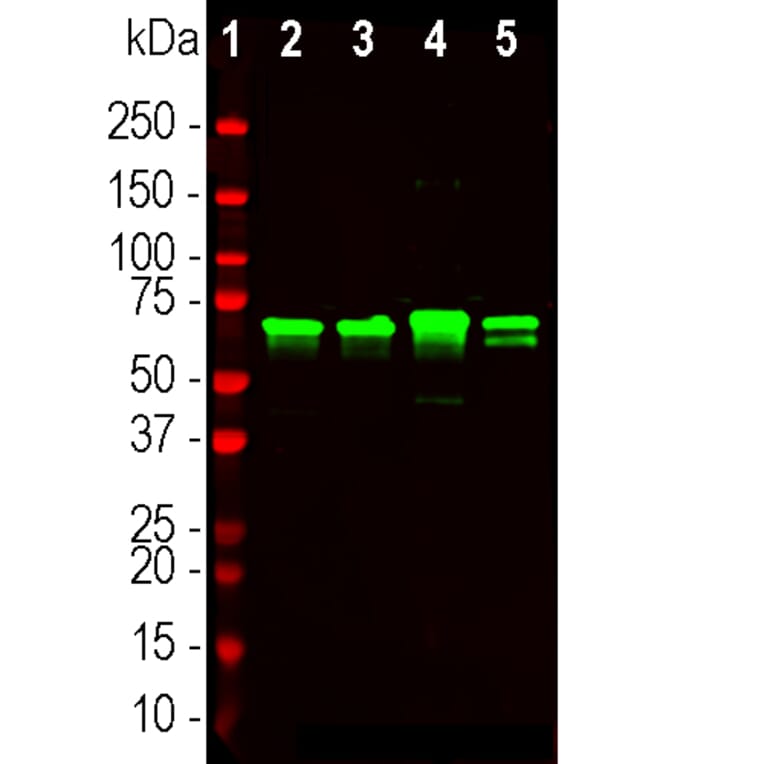
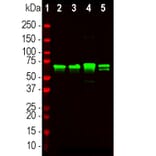
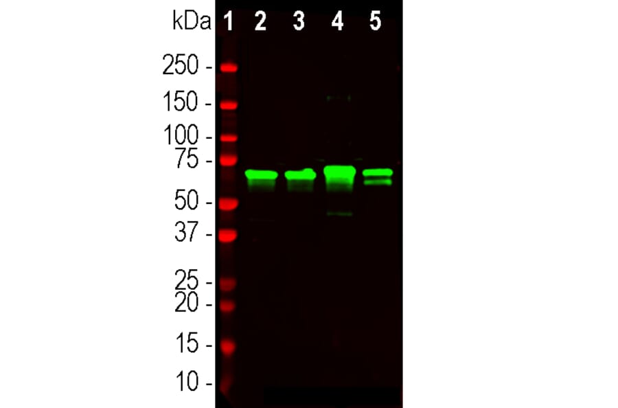
Western blot analysis of different tissue lysates using Anti-NF-L Antibody (A270580), at a dilution of 1:20,000, in green. The lanes contain: [Lane 1] protein standard (red), [Lane 2] rat brain, [Lane 3] rat spinal cord, [Lane 4] mouse brain, and [Lane 5] mouse spinal cord. The strong band at 68 kDa corresponds to the NF-L protein.
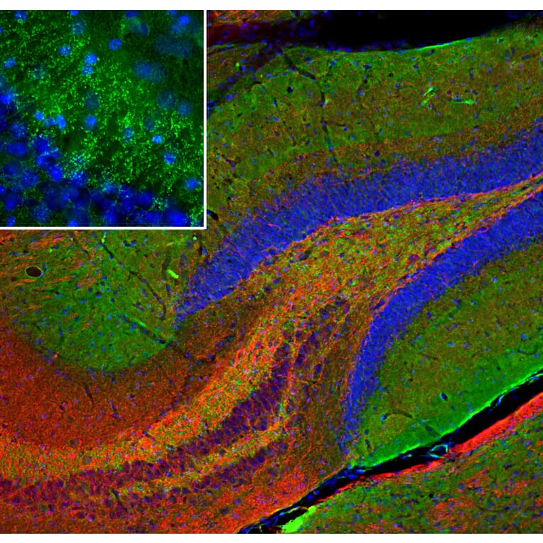
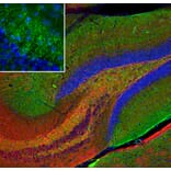
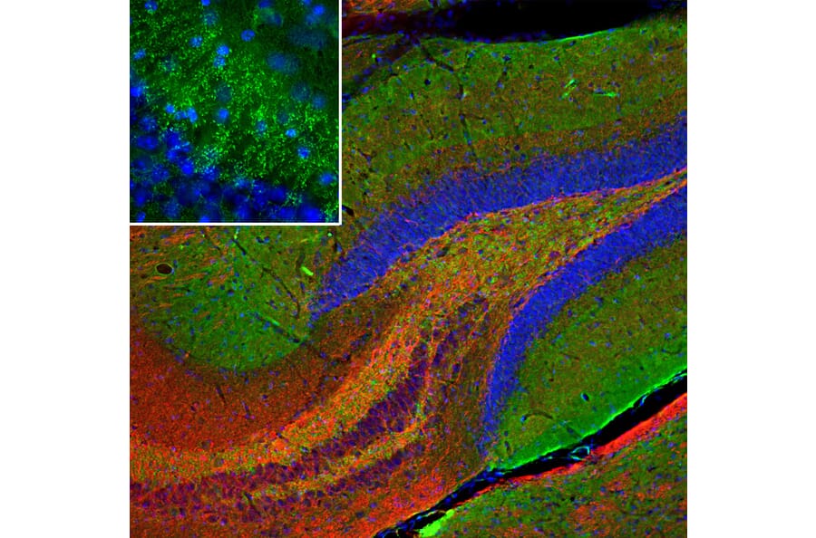
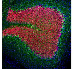
Immunofluorescent analysis of mouse hippocampus section stained with Anti-beta Synuclein Antibody [6A10] (A270558), at a dilution of 1:500, in green, and co-stained with Anti-NF-L Antibody (A270580), at a dilution of 1:5,000, in red. Nuclear DNA is visualised in blue using Hoechst staining. Following transcardial perfusion with 4% paraformaldehyde, the brain was post-fixed for 24 hours, cut to 45 µm, and free-floating sections were stained using the above antibodies. Anti-beta Synuclein Antibody [6A10] (A270558) detects protein concentrated in synaptic regions, while Anti-NF-L Antibody (A270580) labels dendrites and axons of neuronal cells.
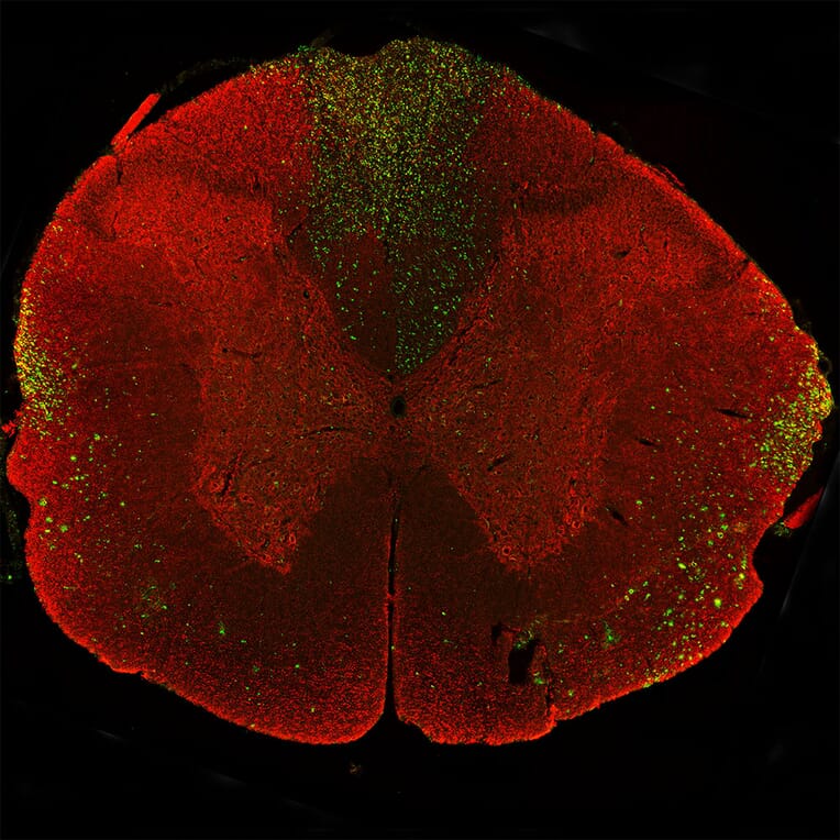
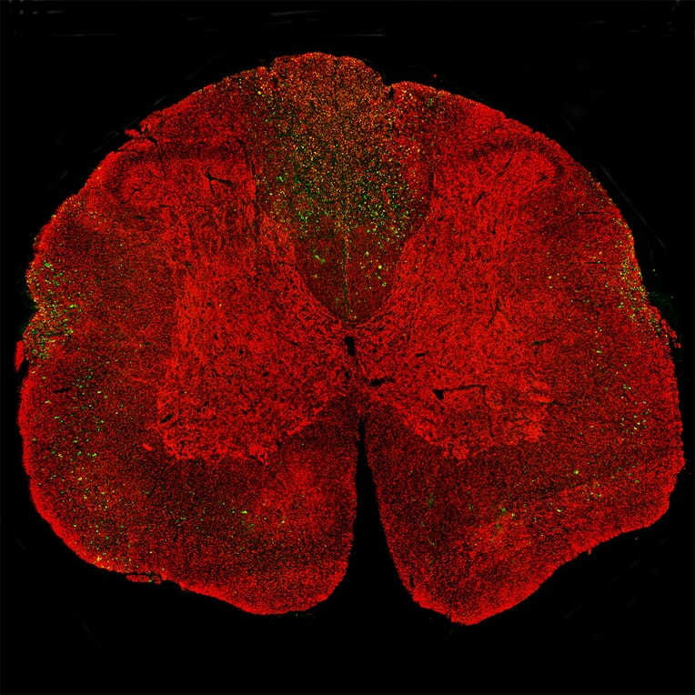
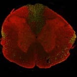
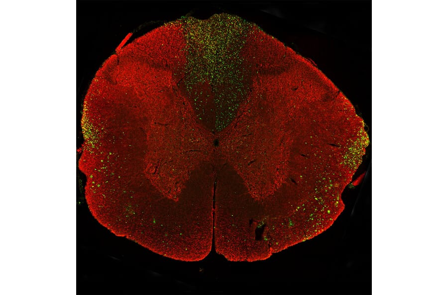
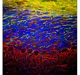
Immunostaining of a coronal section of the spinal cord of a rat given a midline C4 contusion injury three days previously. Sections were stained with Anti-NF-L Antibody (A270580) (red) and Anti-NF-L Antibody [6H63] (A333276) (green). Anti-NF-L Antibody [6H63] (A333276) stains prominent aggregates of material concentrated in the lateral funiculi and the dorsal columns but seen in lesser amounts throughout the section. These are degenerating and degenerated axons damaged by the C4 lesion. The Anti-NF-L Antibody antibody binds the C-terminal “tail” region of NF-L which is absent or destroyed during degeneration, so the positive profiles are largely negative for Anti-NF-L Antibody.
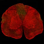
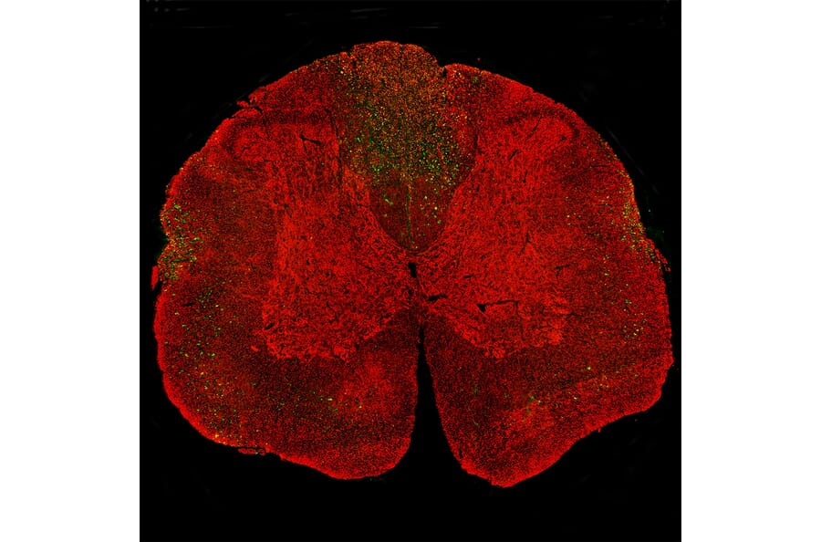
Immunostaining of a coronal section of the spinal cord of a rat given a midline C4 contusion injury three days previously. Sections were stained with Anti-NF-L Antibody (A270580) (red) and Anti-NF-L Antibody [1B11] (A104310) (green). Anti-NF-L Antibody [1B11] (A104310) stains prominent aggregates of material concentrated in the lateral funiculi and the dorsal columns but seen in lesser amounts throughout the section. These are degenerating and degenerated axons damaged by the C4 lesion. The Anti-NF-L Antibody antibody binds the C-terminal “tail” region of NF-L which is absent or destroyed during degeneration, so the positive profiles are largely negative for Anti-NF-L Antibody.
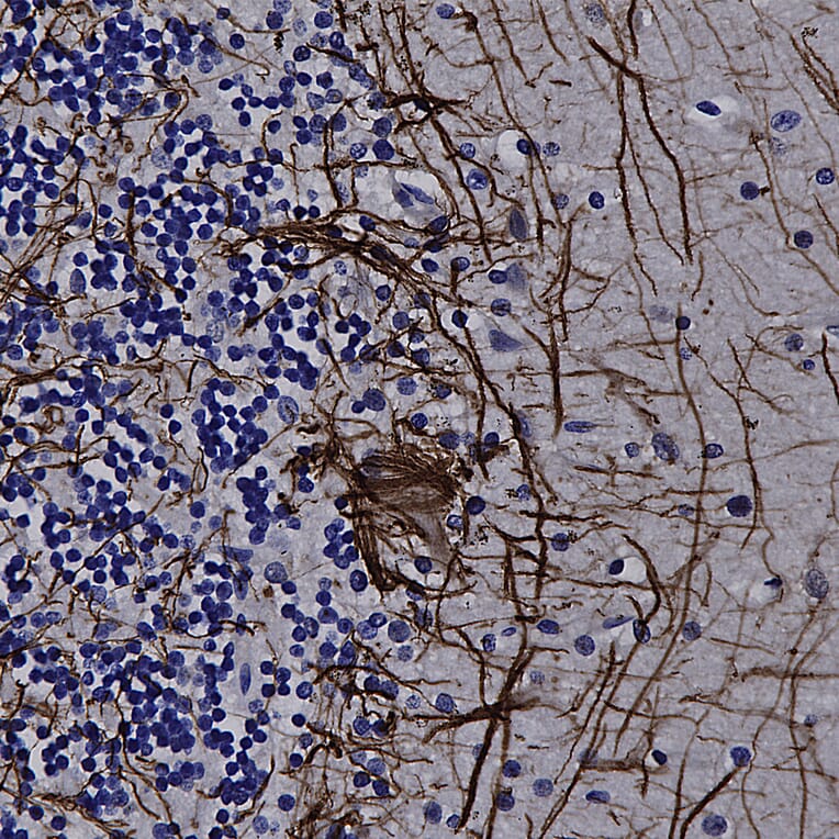
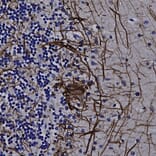
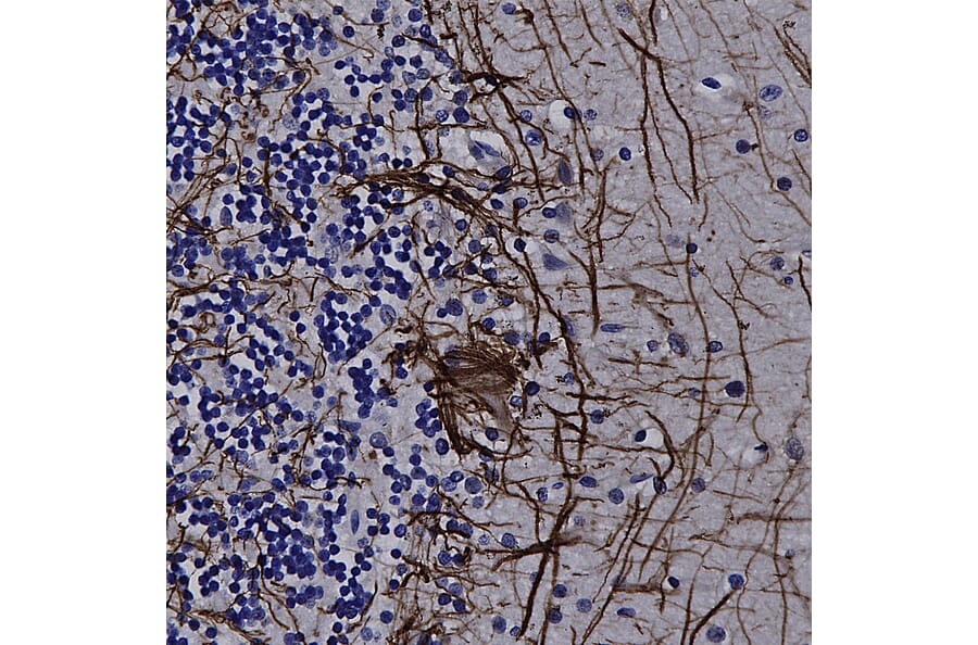
Immunohistochemistry analysis of a formalin fixed paraffin embedded human cerebellum section with Anti-NF-L Antibody (A270580) at a dilution of 1:5,000 detected with DAB (brown) using the Vector Labs ImmPRESS method and reagents with citra buffer retrieval. Counterstained with Hematoxylin (blue). Anti-NF-L Antibody (A270580) strongly labels the axons and dendrites of Purkinje cells and the projections of neuronal cells within the granular layer. Note: this antibody performs well in testing with both 4% PFA and standard NBF fixed rat, mouse, and human tissues.


![Immunofluorescence - Anti-NF-L Antibody [DA2] (A85454) - Antibodies.com](https://cdn.antibodies.com/image/catalog/85/A85454_1.jpg?profile=product_alternative)

![Immunofluorescence - Anti-NF-L Antibody [1B11] (A104310) - Antibodies.com](https://cdn.antibodies.com/image/catalog/104/A104310_1.jpg?profile=product_alternative)
![Immunofluorescence - Anti-NF-L Antibody [7D1] (A85453) - Antibodies.com](https://cdn.antibodies.com/image/catalog/85/A85453_2.jpg?profile=product_alternative)
![Immunohistochemistry - Anti-NF-L Antibody [NR-4] (A252677) - Antibodies.com](https://cdn.antibodies.com/image/catalog/249/A249494_1.jpg?profile=product_alternative)
![Immunohistochemistry - Anti-NF-L Antibody [NR-4] - BSA and Azide free (A249494) - Antibodies.com](https://cdn.antibodies.com/image/catalog/252/A252674_1.jpg?profile=product_alternative)

![Immunohistochemistry - Anti-NF-L Antibody [NFL/736] - BSA and Azide free (A249496) - Antibodies.com](https://cdn.antibodies.com/image/catalog/252/A252676_1.jpg?profile=product_alternative)
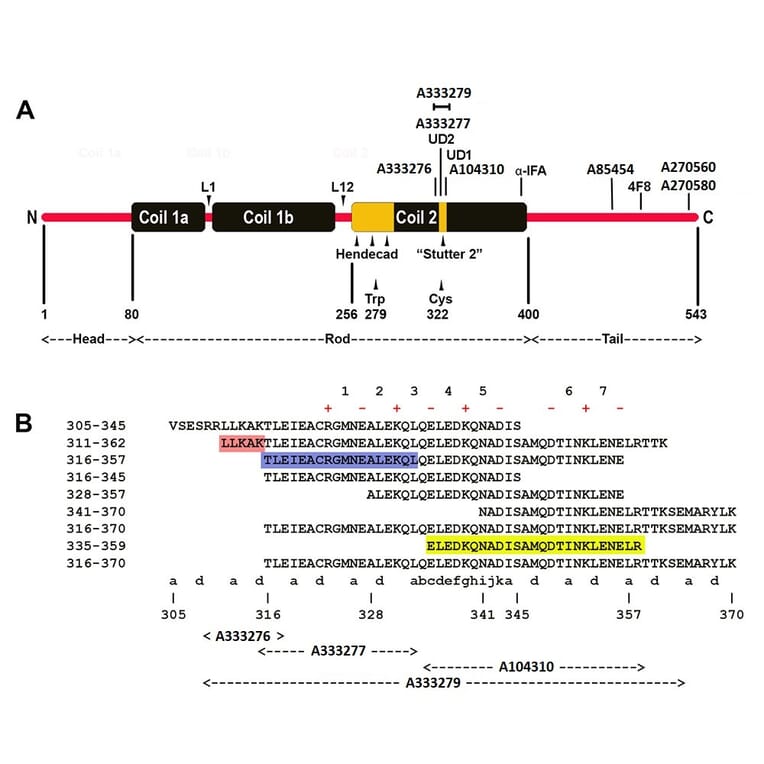
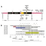
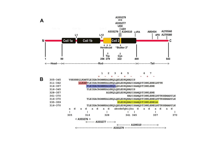
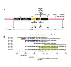
![Epitope Diagram - Anti-NF-L Antibody [6H112] (A270560) - Antibodies.com](https://cdn.antibodies.com/image/catalog/270/A270560_1.jpg?profile=product_alternative)