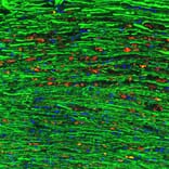
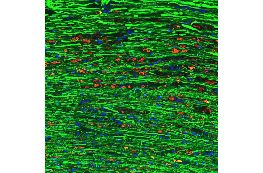
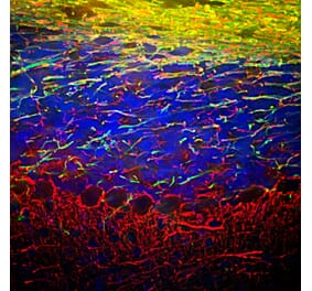
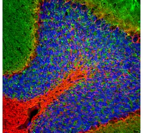
Immunofluorescence of a section of spinal cord from a rat given a C4 contusion injury 3 days previously. The section was stained with Anti-NF-M Antibody [3H11] (A85325) (green) and counterstained with Anti-NF L Antibody (A333279) at a dilution of 1:1,000 (red). The Anti-NF L Antibody (A333279) does not stain the undamaged axons which are strongly positive for the NF-M antibody. However, linear arrays of swollen profiles which originated from damaged axons are strongly positive for the Anti-NF L Antibody (A333279) antibody but not the NF-M antibody, although there is clearly some staining. The Anti-NF-M Antibody [3H11] (A85325) epitope, which is in the C-terminal “tail” of NF-M, has either been partially removed or destroyed.
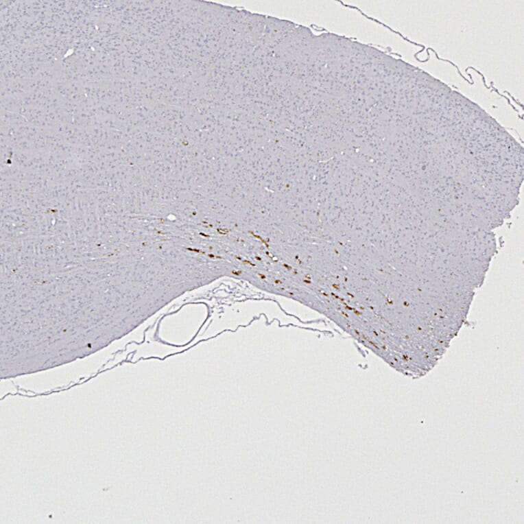
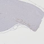
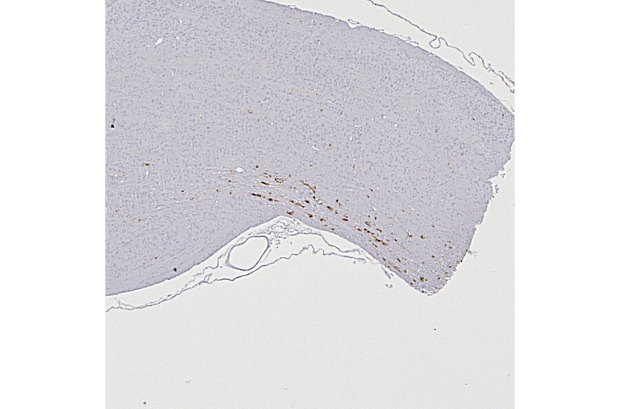
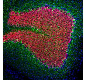
Immunohistochemistry analysis of formalin fixed paraffin embedded heterozygous G85R-SOD1-YFP mice injected with Anti-NF L Antibody (A333279) at a dilution of 1:5,000 detected with DAB (brown) using the Vector Labs ImmPRESS method and reagents with no antigen retrieval. Counterstained with Hematoxylin (blue).
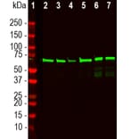
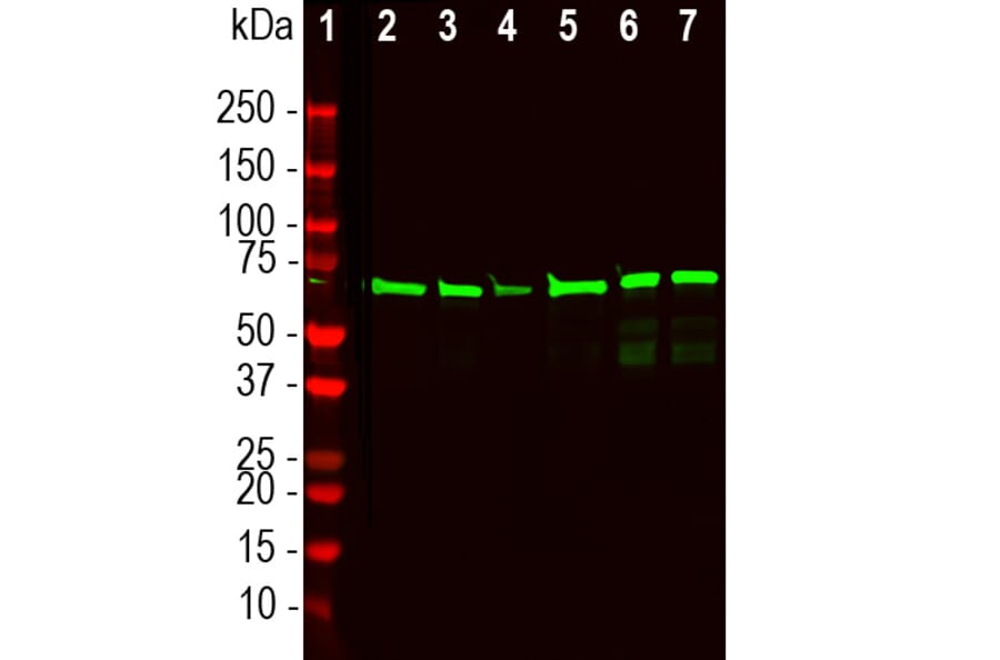
Western blot analysis of different tissue lysates using Anti-NF L Antibody (A333279) at a dilution of 1:2,000 (green). The lanes contain: [Lane 1] protein standard, [Lane 2] rat brain, [Lane 3] rat spinal cord, [Lane 4] mouse brain, [Lane 5] mouse spinal cord, [Lane 6] cow spinal cord, and [Lane 7] pig spinal cord. The strong band at about 68kDa corresponds to full length denatured NF-L protein.
Publishing research using Anti-NF-L Antibody (A333279)? Please let us know so that we can list the citation on this page.
Alternative products to Anti-NF-L Antibody (A333279)


![Immunofluorescence - Anti-NF-L Antibody [DA2] (A85454) - Antibodies.com](https://cdn.antibodies.com/image/catalog/85/A85454_1.jpg?profile=product_alternative)
![Immunofluorescence - Anti-NF-L Antibody [1B11] (A104310) - Antibodies.com](https://cdn.antibodies.com/image/catalog/104/A104310_1.jpg?profile=product_alternative)
![Immunofluorescence - Anti-NF-L Antibody [7D1] (A85453) - Antibodies.com](https://cdn.antibodies.com/image/catalog/85/A85453_2.jpg?profile=product_alternative)
![Immunohistochemistry - Anti-NF-L Antibody [NR-4] (A252677) - Antibodies.com](https://cdn.antibodies.com/image/catalog/249/A249494_1.jpg?profile=product_alternative)
![Immunohistochemistry - Anti-NF-L Antibody [NR-4] - BSA and Azide free (A249494) - Antibodies.com](https://cdn.antibodies.com/image/catalog/252/A252674_1.jpg?profile=product_alternative)

![Immunohistochemistry - Anti-NF-L Antibody [NFL/736] - BSA and Azide free (A249496) - Antibodies.com](https://cdn.antibodies.com/image/catalog/252/A252676_1.jpg?profile=product_alternative)
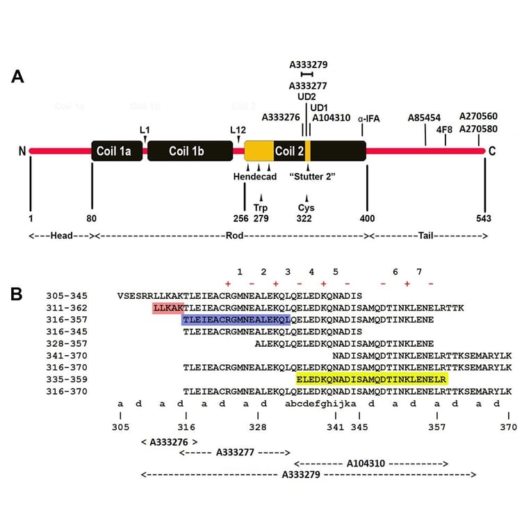
![Epitope Diagram - Anti-NF-L Antibody [6H112] (A270560) - Antibodies.com](https://cdn.antibodies.com/image/catalog/270/A270560_1.jpg?profile=product_alternative)