The epitope of this antibody is not fully characterized, since it could not be localized using 20 amino acid nested peptides, however, it is known to be not within the a-helical region, amino acids 88-400, of the human protein.
Applications
WB, ICC/IF, IHC
Dilutions
WB: 1:5,000-1:10,000, ICC/IF: 1:100-1:500
Reactivity
Human, Horse, Cow, Porcine, Rat, Mouse
Immunogen
Full-length native NF-L protein purified from pig spinal cord.
Host
Mouse
Clonality
Monoclonal
Clone ID
7D1
Isotype
IgG2b
Light Chains
kappa
Conjugate
Unconjugated
Purification
Immunogen affinity purification.
Concentration
1 mg/ml
Molecular Weight
68-70 kDa
Product Form
Liquid
Formulation
Supplied in Phosphate Buffered Saline with 50% Glycerol and 5mM Sodium Azide.
Storage
Shipped at 4°C. Upon delivery aliquot and store at -20°C. Avoid freeze/thaw cycles.
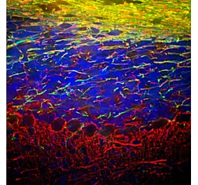
Embryonic rat cortical cells grown in tissue culture and stained with Anti-NF-L Antibody (green) and Anti-NF-H Antibody (A85337 | red). The perikaryal and dendritic neurofilaments in the Large cell in middle are stained with Anti-NF-L Antibody but not with Anti-NF-H Antibody. In contrast both antibodies stain axonal neurofilaments which therefore appear orange.
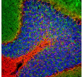
Immunofluorescent analysis of a section of mouse cerebellum stained with Anti-NF-L Antibody [7D1] (A85453), at a dilution of 1:5,000 in green, and co-stained with Anti-FOX2 Antibody (A104328), at a dilution of 1:2,000 in red. The nuclear DNA is visualised in blue using Hoechst staining. Following transcardial perfusion of the mouse with 4% paraformaldehyde, the brain was post-fixed for 24 hours, cut to 45 µm, and free-floating sections were stained with the above antibodies. Anti-NF-L Antibody [7D1] (A85453) labels dendrites and axons of neuronal cells predominantly axons in white matter and basket cell axons associated with Purkinje cells. The Anti-FOX2 Antibody (A104328) reveals protein expressed in a subset of neurons, including the Purkinje cells.
Western blot analysis of different tissue lysates using Anti-NF-L Antibody [7D1] (A85453), at a dilution of 1:5,000, in green. The lanes contain samples of: [Lane 1] Protein standards, in red, [Lane 2] rat brain, [Lane 3] rat spinal cord, [Lane 4] mouse brain, and [Lane 5] mouse spinal cord. The strong band at 68 kDa corresponds to the NF-L protein.
Strip blots of rat spinal cord homogenate probed with two subclones of Anti-NF-L Antibody. Both subclones, one of which became the definitive antibody, stain strongly and specifically a band at 68 kDa with essentially no background.
Publishing research using Anti-NF-L Antibody [7D1] (A85453)? Please let us know so that we can list the citation on this page.
Alternative products to Anti-NF-L Antibody [7D1] (A85453)


![Immunofluorescence - Anti-NF-L Antibody [7D1] (A85453) - Antibodies.com](https://cdn.antibodies.com/image/catalog/85/A85453_2.jpg?profile=product_top)
![Immunofluorescence - Anti-NF-L Antibody [7D1] (A85453) - Antibodies.com](https://cdn.antibodies.com/image/catalog/85/A85453_3.jpg?profile=product_top)
![Western Blot - Anti-NF-L Antibody [7D1] (A85453) - Antibodies.com](https://cdn.antibodies.com/image/catalog/85/A85453_4.jpg?profile=product_top)
![Western Blot - Anti-NF-L Antibody [7D1] (A85453) - Antibodies.com](https://cdn.antibodies.com/image/catalog/85/A85453_5.jpg?profile=product_top)
![Immunofluorescence - Anti-NF-L Antibody [7D1] (A85453) - Antibodies.com](https://cdn.antibodies.com/image/catalog/85/A85453_2.jpg?profile=product_top_thumb)
![Immunofluorescence - Anti-NF-L Antibody [7D1] (A85453) - Antibodies.com](https://cdn.antibodies.com/image/catalog/85/A85453_3.jpg?profile=product_top_thumb)
![Western Blot - Anti-NF-L Antibody [7D1] (A85453) - Antibodies.com](https://cdn.antibodies.com/image/catalog/85/A85453_4.jpg?profile=product_top_thumb)
![Western Blot - Anti-NF-L Antibody [7D1] (A85453) - Antibodies.com](https://cdn.antibodies.com/image/catalog/85/A85453_5.jpg?profile=product_top_thumb)
![Immunofluorescence - Anti-NF-L Antibody [7D1] (A85453) - Antibodies.com](https://cdn.antibodies.com/image/catalog/85/A85453_2.jpg?profile=product_image)
![Immunofluorescence - Anti-NF-L Antibody [7D1] (A85453) - Antibodies.com](https://cdn.antibodies.com/image/catalog/85/A85453_3.jpg?profile=product_image)
![Western Blot - Anti-NF-L Antibody [7D1] (A85453) - Antibodies.com](https://cdn.antibodies.com/image/catalog/85/A85453_4.jpg?profile=product_image)
![Western Blot - Anti-NF-L Antibody [7D1] (A85453) - Antibodies.com](https://cdn.antibodies.com/image/catalog/85/A85453_5.jpg?profile=product_image)
![Immunofluorescence - Anti-NF-L Antibody [DA2] (A85454) - Antibodies.com](https://cdn.antibodies.com/image/catalog/85/A85454_1.jpg?profile=product_alternative)
![Immunofluorescence - Anti-NF-L Antibody [1B11] (A104310) - Antibodies.com](https://cdn.antibodies.com/image/catalog/104/A104310_1.jpg?profile=product_alternative)

![Immunohistochemistry - Anti-NF-L Antibody [NR-4] (A252677) - Antibodies.com](https://cdn.antibodies.com/image/catalog/249/A249494_1.jpg?profile=product_alternative)
![Immunohistochemistry - Anti-NF-L Antibody [NR-4] - BSA and Azide free (A249494) - Antibodies.com](https://cdn.antibodies.com/image/catalog/252/A252674_1.jpg?profile=product_alternative)

![Immunohistochemistry - Anti-NF-L Antibody [NFL/736] - BSA and Azide free (A249496) - Antibodies.com](https://cdn.antibodies.com/image/catalog/252/A252676_1.jpg?profile=product_alternative)
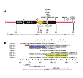
![Epitope Diagram - Anti-NF-L Antibody [6H112] (A270560) - Antibodies.com](https://cdn.antibodies.com/image/catalog/270/A270560_1.jpg?profile=product_alternative)