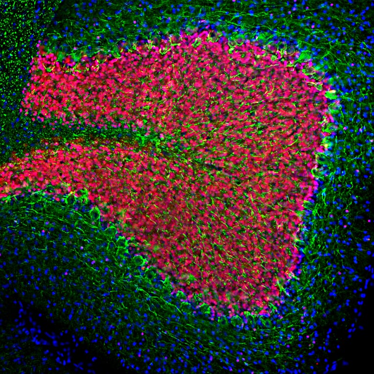
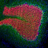
Immunofluorescent analysis of rat cerebellum section stained with Anti-NF-L Antibody (A85286), at a dilution of 1:2,000, in green, and co-stained with Anti-NeuN Antibody [1B7] (A85405), at a dilution of 1:5,000, in red. Following transcardial perfusion with 4% paraformaldehyde, brain was post fixed for 24 hours, cut to 45µM, and free-floating sections were stained with above antibodies. Anti-NF-L Antibody (A85286) labels perikarya and processes of neuronal cells, particularly strongly the axons of basket cells, while Anti-NeuN Antibody [1B7] (A85405) stains the nuclei and proximal cytoplasm of neurons.
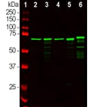
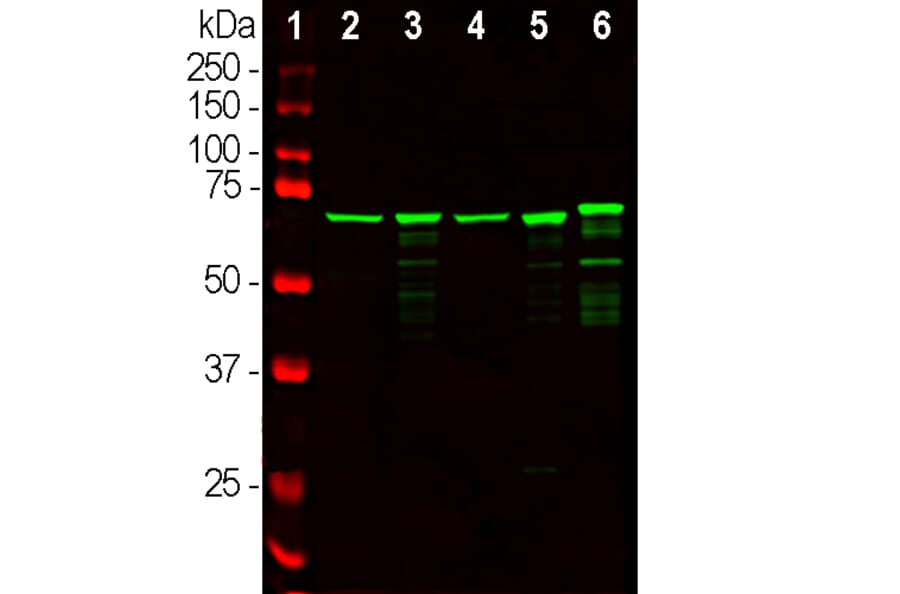
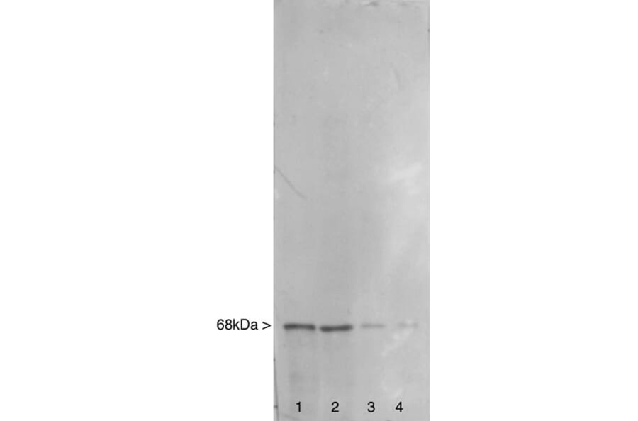
Western blot analysis of different tissue lysates using Anti-NF-L Antibody (A85286), at a dilution of 1:20,000, in green. The lanes contain: [Lane 1] protein standard (red), [Lane 2] rat brain, [Lane 3] rat spinal cord, [Lane 4] mouse brain, [Lane 5] mouse spinal cord, and [Lane 6] cow spinal cord. Strong bands at ~68kDa correspond to NF-L proteins; which are known to have slightly different apparent SDS-PAGE molecular weights across species boundaries.
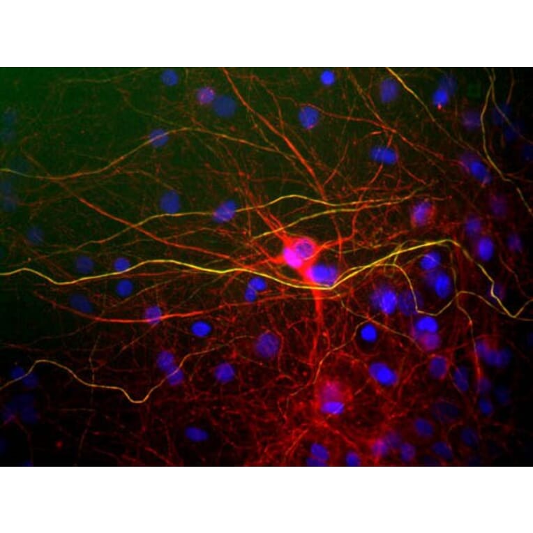
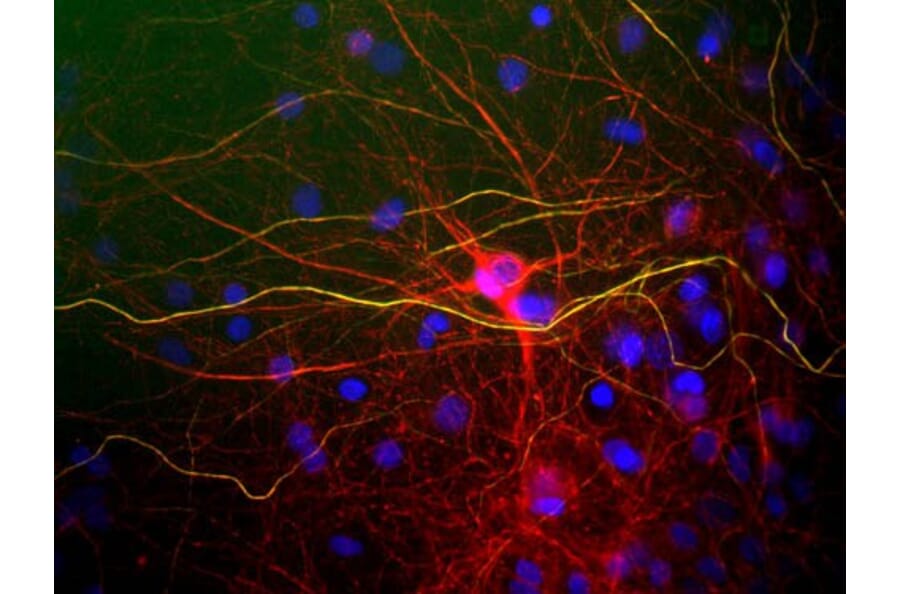
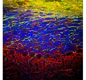
Immunofluorescent analysis of mixed neuron/glial cultures stained with Anti-NF-L Antibody (A85286), in red, and Anti-NF-H Antibody (A85336), in green. The NF-L protein is assembled into neurofilaments which are found throughout the axons, dendrites, and perikarya of these cells. In contrast the phosphorylated NF-H has a more restricted expression pattern, being found only in developed axonal neurofilaments. Axonal profiles therefore are yellow since the red and green signals are superimposed. In contrast neurofilaments containing only NF-L appear red.
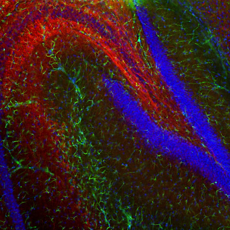
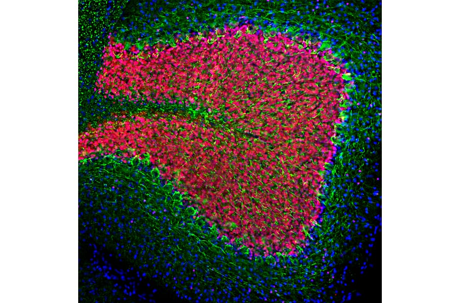
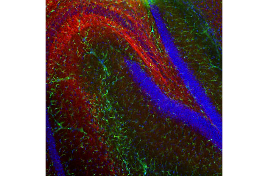
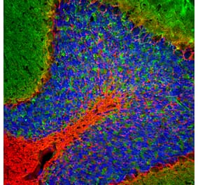
Immunofluorescent analysis of mouse hippocampus section stained with Anti-GFAP Antibody [3E10] (A270551) at a dilution of 1:500 (green) and costained with Anti-NF-L Antibody (A85286) at a dilution of 1:2,000 (red). Nuclei were stained with Hoechst (blue). Following transcardial perfusion of mouse with 4% paraformaldehyde, brain was post fixed for 24 hours, cut to 45µM, and free-floating sections were stained with the above antibodies. The GFAP antibody stains network of glial cells while the Anti-NF-L Antibody antibody labels axons and dendrites of neuronal cells.
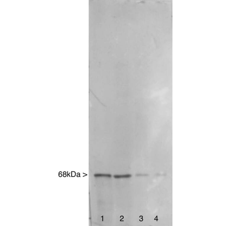
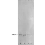
Western blot analysis of different tissue lysates using Anti-NF-L Antibody (A85286). The lanes contain: [Lane 1] rat spinal cord, [Lane 2] rat brain stem, [Lane 3] rat cerebellum, and [Lane 4] rat cerebral cortex. Neurofilaments are concentrated in large projection axons and therefore NF-L is a more prominent component of spinal cord than cortical regions.
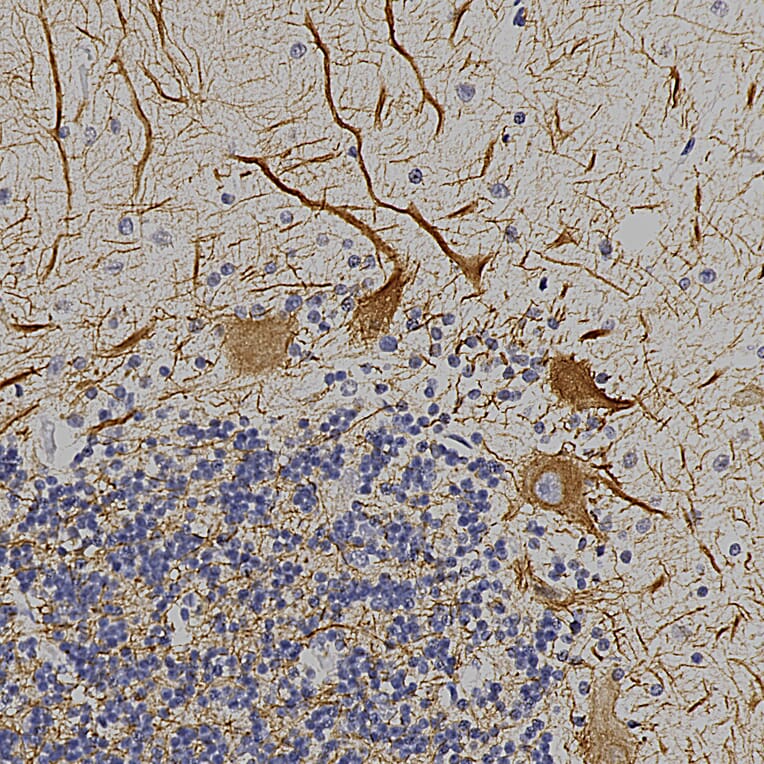
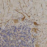
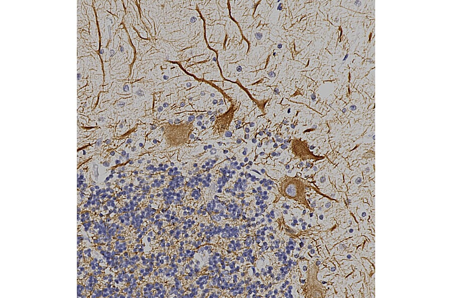
Immunohistochemistry analysis of a formalin fixed paraffin embedded human cerebellum with Anti-NF-L Antibody (A85286) at a dilution of 1:4,000 detected in DAB (brown) following the ABC method. Counterstained with Hematoxylin (blue). The NF-L antibody detects perikaryal and dendritic processes of Purkinje cells and axons of other neuronal cells, particularly basket cells.


![Immunofluorescence - Anti-NF-L Antibody [DA2] (A85454) - Antibodies.com](https://cdn.antibodies.com/image/catalog/85/A85454_1.jpg?profile=product_alternative)
![Immunofluorescence - Anti-NF-L Antibody [1B11] (A104310) - Antibodies.com](https://cdn.antibodies.com/image/catalog/104/A104310_1.jpg?profile=product_alternative)

![Immunofluorescence - Anti-NF-L Antibody [7D1] (A85453) - Antibodies.com](https://cdn.antibodies.com/image/catalog/85/A85453_2.jpg?profile=product_alternative)
![Immunohistochemistry - Anti-NF-L Antibody [NR-4] (A252677) - Antibodies.com](https://cdn.antibodies.com/image/catalog/249/A249494_1.jpg?profile=product_alternative)

![Immunohistochemistry - Anti-NF-L Antibody [NR-4] - BSA and Azide free (A249494) - Antibodies.com](https://cdn.antibodies.com/image/catalog/252/A252674_1.jpg?profile=product_alternative)
![Immunohistochemistry - Anti-NF-L Antibody [NFL/736] - BSA and Azide free (A249496) - Antibodies.com](https://cdn.antibodies.com/image/catalog/252/A252676_1.jpg?profile=product_alternative)
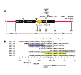
![Epitope Diagram - Anti-NF-L Antibody [6H112] (A270560) - Antibodies.com](https://cdn.antibodies.com/image/catalog/270/A270560_1.jpg?profile=product_alternative)