Claudie Hooper, PhD | 13th April 2023
Epitope tags are short amino acid sequences that are genetically engineered to be expressed in-frame at the beginning or end of a protein (at the N or C terminus) without altering the structure, sub-cellular localisation or function of the native protein. Epitope tagging enables the subsequent detection of the protein of interest using a corresponding specific epitope tag antibody. This is achieved through expressing epitope tagged proteins in host cells using an expression vector into which the tagged protein has been previously cloned.
Epitope tagging serves as a useful technique to enable the detection of proteins of low abundance, low immunogenicity, or proteins where commercial antibodies are not yet available. Antibodies raised against epitope tags can be used experimentally in a variety of applications such as western blotting, immunoprecipitation, co-immunoprecipitation for the investigation of protein-protein interactions, flow cytometry, immunofluorescence, immunocytochemistry, and immunohistochemistry. Note in western blotting the epitope tag causes a shift in the molecular weight of the protein in accordance with the length of the epitope tag.
There are many epitope tags available and the choice of epitope tag depends on the intended downstream application to analyze the tagged protein. Epitope tags can be divided into three categories: Affinity Tags, such as His tag, Glutathione S-Transferase (GST), and Maltose Binding Protein (MBP), are commonly used to purify recombinant proteins; Fluorescent Protein Tags, such as Green Fluorescent Protein (GFP), Red Fluorescent Protein (RFP), mCherry, Fluorescein Isothiocyanate (FITC), and tdTomato, can be used for cell tracking, sub-cellular localisation and cell sorting; and Peptide Tags, such as Hemagglutinin (HA) Tag, c-Myc, V5 Tag, and FLAG Tag, may be used for protein detection and the investigation of protein-protein interactions.
| Epitope Tag | Amino Acids | Amino Acid Sequence |
|---|---|---|
| Avi Tag | 15 | GLNDIFEAQKIEWHE |
| Myc Tag | 10 | EQKLISEEDL |
| E Tag | 13 | GAPVPYPDPLEPR |
| FLAG Tag | 8 | DYKDDDDK |
| HA Tag | 9 | YPYDVPDYA |
| His Tag | 6 | HHHHHH |
| HSV Tag | 11 | QPELAPEDPED |
| S1 Tag | 9 | NANNPDWDF |
| Strep II Tag | 9 | NWSHPQFEK |
| T7 Tag | 11 | MASMTGGQQMG |
| TAP Tag | 21 | CSSGALDYDIPTTASENLYFQ |
| V5 Tag | 14 | GKPIPNPLLGLDST |
| VSV-G Tag | 11 | YTDIEMNRLGK |
| X-press Tag | 8 | DLYDDDDK |
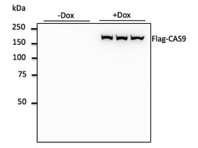
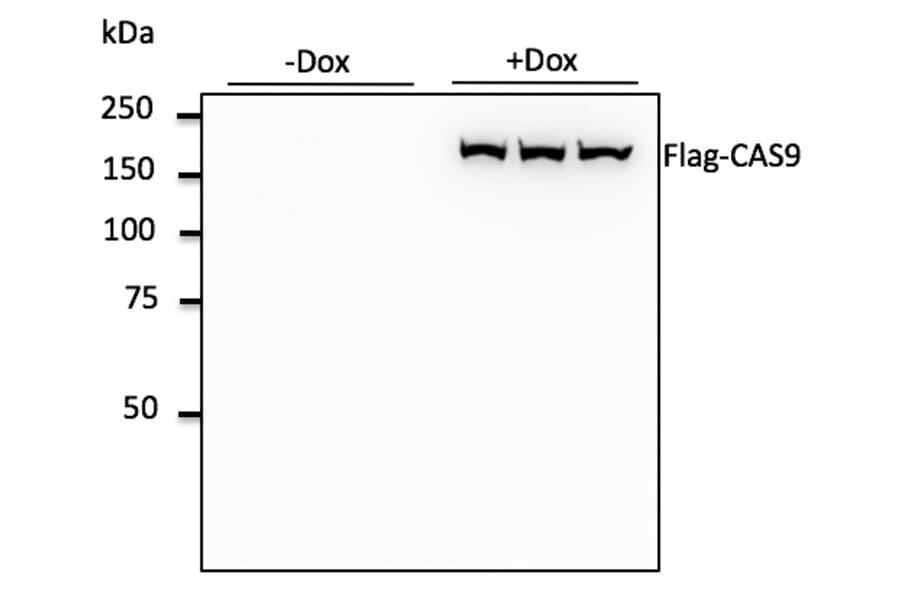
Figure 1: 293 hTERT RPE-1 cells, transduced with Flag-CAS9 lentivirus, detected with Anti-FLAG Tag Antibody (A121606) at a 1:1,000 dilution. Lysates at 50µg per lane and rabbit anti-goat IgG antibody (HRP) at a 1:10,000 dilution.
Figure 2: Immunoprecipitation of 293T cells expressing a His Tag fusion protein by Anti-His Tag Antibody [RM146] (A121323), and then blotted with Anti-His Tag Antibody [RM146]. (1) Whole lysate control. (2) IP by rabbit IgG control. (3) IP by Anti-His Tag Antibody [RM146].
The HA epitope tag originates from amino acids 98-106 of human influenza HA, which is a protein involved in viral infectivity.
| Recommended Antibodies | Validated Applications |
|---|---|
| Anti-HA Tag Antibody (A121677) | WB |
| Anti-HA Tag Antibody [HA.C5] (A85278) | Dot Blot, ELISA, IP, WB |
| Anti-HA Tag Antibody [16.43] (A250863) | ELISA, IF, WB, IP, Flow Cytometry, IHC-P |
His tag, also known as the polyhistidine tag, is an artificial peptide consisting of six to nine histidine residues.
| Recommended Antibodies | Validated Applications |
|---|---|
| Anti-His Tag Antibody [HIS.H8] (A85277) | Dot Blot, ELISA, IP, WB |
| Recombinant Anti-His Tag Antibody [RM146] (A121323) | WB, IP, ICC, Flow Cytometry, IHC |
The FLAG tag, otherwise known as the DYKDDDDK tag, is an artificial octapeptide that is not derived from a naturally occurring protein. This tag contains a recognition sequence for the protease Enterokinase, enabling cleavage of FLAG-tagged proteins and thus tag removal.
| Recommended Antibodies | Validated Applications |
|---|---|
| Anti-FLAG Tag Antibody (A121606) | WB |
| Anti-FLAG Tag Antibody [F-tag-01] (A85906) | WB, ICC |
| Anti-FLAG Tag Antibody [FG4R] (A85282) | Dot Blot, ELISA, IP, WB |
The Myc epitope tag is derived from the C-terminal, amino acids 410-419, of the c-Myc protein, which is a transcription factor involved in cell division.
| Recommended Antibodies | Validated Applications |
|---|---|
| Anti-c-Myc Antibody [9E10] (A85464) | Flow Cytometry, IP, WB, IHC-P |
| Anti-c-Myc Antibody [9E10] (HRP) (A85463) | WB |
| Anti-Myc Tag Antibody [MYC.A7] (A85281) | Dot Blot, ELISA, IP, WB |
The T7 epitope tag is a peptide of 11 amino acids encoded in the leader sequence of the T7 bacteriophage gene 10, which encodes a T7 major capsid protein whose exact function remains to be elucidated.
| Recommended Antibody | Validated Applications |
|---|---|
| Anti-T7 Tag Antibody (A121634) | WB |
The V5 epitope tag is derived from an epitope (either 14 or 9 amino acids) present on the P and V proteins of the paramyxovirus of the Simian Virus 5 family.
| Recommended Antibodies | Validated Applications |
|---|---|
| Anti-V5 Tag Antibody [V5.E10] (A85280) | Dot Blot, ELISA, IP, WB |
| Anti-V5 Tag Antibody (A121614) | WB, IF |
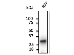
Fluorescent protein tags represent another useful tool that can be used in a variety of laboratory applications including cell tracking, sub-cellular localisation and cell sorting. Commonly used fluorescent protein tags include Green Fluorescent Protein (GFP) - derived from the jellyfish Aequorea victoria - and Red Fluorescent Protein (RFP). RFP is a variant of DsRed - originally isolated from the sea anemone, Discosoma sp, based on its homology to jellyfish GFP – and other common variants include mCherry, mPlum, and tdTomato. We offer a comphrensive range of fluorescent protein tag antibodies, including antibodies against DENDRA2, FITC, GFP, HRP, mCherry, mPlum, RFP, and tdTomato.
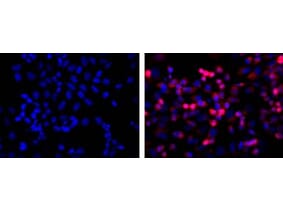
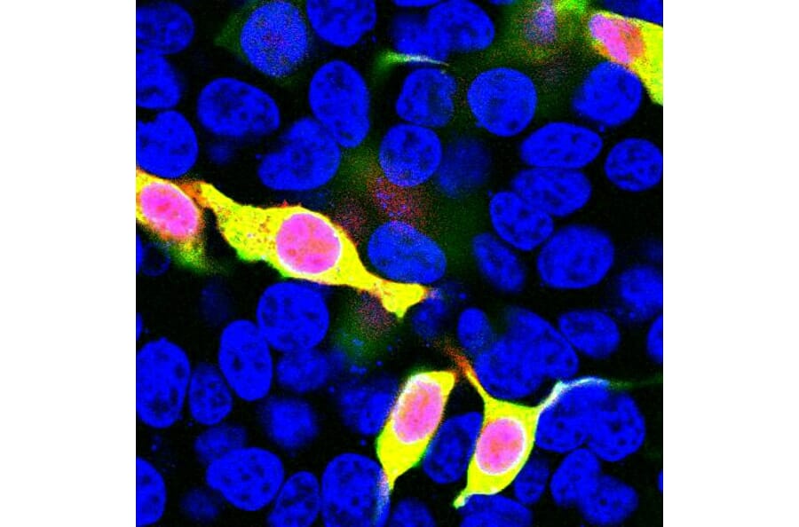
Figure 3: Immunofluorescent analysis of transfected HEK293 cells with a mCherry-HA construct, in red, and stained with Anti-mCherry Antibody (A85306), dilution 1:1,000, in green. The blue is Hoechst staining of nuclear DNA. Anti-mCherry Antibody (A85306) reveals mCherry protein expressed only in transfected cells which appear golden in color.
Figure 4: Immunofluorescent analysis of transfected HEK293 cells with a GFP construct, in green, and stained with Anti-GFP Antibody (A85298), dilution 1:2,000, in red. The blue is Hoechst staining of nuclear DNA. Anti-GFP Antibody (A85298) reveals GFP protein expressed only in transfected cells, and as a result these cells appear orange-yellow in color.
| Fluorescent Protein Tag Antibody | Validated Applications |
|---|---|
| Anti-DENDRA2 Antibody (A121799) | WB, ICC, Flow Cytometry |
| Anti-FITC Antibody [SPM395] (A250900) | Flow Cytometry, IF, WB, IHC-P |
| Anti-GFP Antibody (A290) | ELISA, IHC-Fr, ICC, IHC-P, IP, WB, IHC-FoFr, IHC-FrFl |
| Anti-HRP Antibody [HP-03] (A86768) | WB, ICC, ELISA |
| Anti-mCherry Antibody (A85306) | WB, ICC/IF, IHC |
| Anti-mPlum Antibody (A285875) | WB, ICC, Flow Cytometry |
| Anti-RFP Antibody (A121675) | WB, IF, IHC-P, IHC-Fr, IEM |
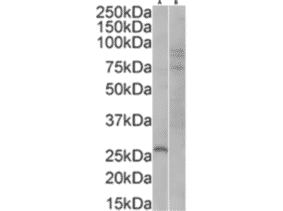
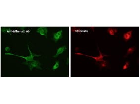
| Anti-tdTomato Antibody (A121690) | WB, IF, IHC-P, IHC-Fr, IEM |
![Immunoprecipitation - Anti-His Tag Antibody [RM146] (A121323) - Antibodies.com](https://cdn.antibodies.com/image/catalog/121/A121323_2.png?profile=product_image)

![Anti-HA Tag Antibody [HA.C5] - A85278 - Antibodies.com](https://cdn.antibodies.com/image/resources/A85278_Popular.png?profile=product_alternative_3)
![Anti-mCherry Antibody [1C51] - A85305 - Antibodies.com](https://cdn.antibodies.com/image/resources/A85305_Popular.png?profile=product_alternative_3)
![Western Blot - Anti-HRP Antibody [HP-03] (A86767) - Antibodies.com](https://cdn.antibodies.com/image/catalog/86/A86768_876.jpg?profile=product_alternative_3)
![Western Blot - Anti-GST Tag Antibody [S-tag-05] (A85569) - Antibodies.com](https://cdn.antibodies.com/image/catalog/85/A85570_72.jpg?profile=product_alternative_3)