

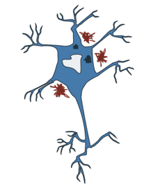
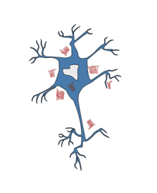
Neurodegeneration is the progressive loss of neurons via apoptosis, oxidative stress, structural abnormalities, or a general failure to function. It can take place at the molecular level or be part of a larger, systemic failure within the nervous system. Humans are now living longer than ever before and, as a result, disease associated with ageing is becoming increasingly prevalent. A rapidly ageing society has resulted in neurodegenerative conditions such as Alzheimer’s disease, post-stroke dementia, and Parkinson’s disease becoming a significant problem. Consequently, neurodegenerative disease has become one of the most exciting and well researched areas of neuroscience.
The exact mechanisms, pathology, and causes behind neurodegeneration is varied, complex, and largely unknown but can range from protein misfolding, dysfunction of intracellular mechanism, genetics, mitochondrial dysfunction and oxidative stress, structural damage, and programmed cell death / apoptosis. Prion diseases are the exception, as these can be either geneticly determined or infectiously spread. Neurodegenerative diseases are currently incurable, however, advances in research show that there are many similarities between these diseases on a sub-cellular level; including atypical protein assemblies and induced cell death. This discovery provides the hope of therapeutic advances that could treat multiple diseases simultaneously.
We offer a range of antibodies against markers and related proteins for Alzheimers' disease, amyotrophic lateral sclerosis, Parkinson's disease, Huntington's disease, and prion diseases, displayed below.
Alzheimer’s disease is a chronic, progressive neurodegenerative disease. It is estimated to be the cause of around 60-70% of all dementia cases; affecting approximately twelve million individuals worldwide. It is initially characterized by difficulty remembering recent events, with progression of the disease indicated by difficulty with speech, disorientation, mood swings, and other behavioural issues. Due to the gradual onset of the symptoms, it can be many years before a diagnosis is formally made. Though the exact cause of the disease is unknown, it is associated with hallmark extracellular amyloid-β plaques and intracellular tau-based neurofibrillary tangles; which are thought to potentially contribute to the development of the disease.
Amyloid build up is formed from the peptide cleavage products of the amyloid beta precursor protein (APP), with tau caused by the hyperphosphorylation of tau-based tangles that bind and clump together, and both are thought to disrupt normal neuronal function. Research is currently focused on this amyloid-tau hypothesis. However, several other theories are also being considered, such as neurovascular failure, poor cell homeostasis, or a dysfunctional immune system.
This gene encodes a cell surface receptor and transmembrane precursor protein that is cleaved by secretases to form a number of peptides. Some of these peptides are secreted and can bind to the acetyltransferase complex APBB1/TIP60 to promote transcriptional activation, while others form the protein basis of the amyloid plaques found in the brains of patients with Alzheimer disease. In addition, two of the peptides are antimicrobial peptides, having been shown to have bacteriocidal and antifungal activities. Mutations in this gene have been implicated in autosomal dominant Alzheimer disease and cerebroarterial amyloidosis (cerebral amyloid angiopathy). Multiple transcript variants encoding several different isoforms have been found for this gene. Target information from NCBI Gene ID: 351.
| Product Name | Reactivity | Applications |
|---|---|---|
| Anti-Amyloid Precursor Protein Antibody (A448) | Human, Mouse, Rat | WB, ICC, IHC |
| Anti-Amyloid Precursor Protein Antibody (A449) | Human, Mouse, Rat | WB, ICC |
| Anti-Amyloid Precursor Protein Antibody (A83045) | Mouse, Rat | ELISA, WB |
| Anti-Amyloid Precursor Protein Antibody (A83047) | Human, Mouse | ELISA, WB |
| Anti-APP delta C31 Antibody (A451) | Human, Mouse, Rat | WB, ICC, ELISA |
APOE is an apolipoprotein, a protein associating with lipid particles, that mainly functions in lipoprotein-mediated lipid transport between organs via the plasma and interstitial fluids. APOE is a core component of plasma lipoproteins and is involved in their production, conversion and clearance. Apolipoproteins are amphipathic molecules. As such, APOE associates with chylomicrons, chylomicron remnants, very low density lipoproteins (VLDL) and intermediate density lipoproteins (IDL) but shows a preferential binding to high-density lipoproteins (HDL). It also binds a wide range of cellular receptors including the LDL receptor/LDLR, the LDL receptor-related proteins LRP1, LRP2 and LRP8 and the very low-density lipoprotein receptor/VLDLR that mediate the cellular uptake of the APOE-containing lipoprotein particles. Finally, APOE has also a heparin-binding activity and binds heparan-sulfate proteoglycans on the surface of cells, a property that supports the capture and the receptor-mediated uptake of APOE-containing lipoproteins by cells. By participating in the lipoprotein-mediated distribution of lipids among tissues, APOE plays a critical role in plasma and tissues lipid homeostasis. APOE also plays an important role in lipid transport in the central nervous system, regulating neuron survival and sprouting. The APOE*4 allele (APOE form E4) is genetically associated with the common late onset familial and sporadic forms of Alzheimer disease. Risk for AD increased from 20% to 90% and mean age at onset decreased from 84 to 68 years with increasing number of APOE*4 alleles in 42 families with late onset AD. Thus APOE*4 gene dose is a major risk factor for late onset AD and, in these families, homozygosity for APOE*4 was virtually sufficient to cause AD by age 80. The mechanism by which APOE*4 participates in pathogenesis is not known. Target information from Uniprot accession P02649.
| Product Name | Reactivity | Applications |
|---|---|---|
| Anti-Apolipoprotein E Antibody (A84561) | Human | ELISA, WB, IHC |
| Anti-Apolipoprotein E Antibody (A84074) | Human | ELISA, WB |
| Anti-Apolipoprotein E Antibody (A831) | Human | WB |
This gene encodes a member of the peptidase A1 family of aspartic proteases. Alternative splicing results in multiple transcript variants, at least one of which encodes a preproprotein that is proteolytically processed to generate the mature protease. This transmembrane protease catalyzes the first step in the formation of amyloid beta peptide from amyloid precursor protein. Amyloid beta peptides are the main constituent of amyloid beta plaques, which accumulate in the brains of human Alzheimer's disease patients. Target information from NCBI Gene ID: 23621.
| Product Name | Reactivity | Applications |
|---|---|---|
| Anti-BACE1 Antibody (A43142) | Human | WB |
Sialic-acid-binding immunoglobulin-like lectin (Siglec) that plays a role in mediating cell-cell interactions and in maintaining immune cells in a resting state. Preferentially recognizes and binds alpha-2,3- and more avidly alpha-2,6-linked sialic acid-bearing glycans. Upon engagement of ligands such as C1q or syalylated glycoproteins, two immunoreceptor tyrosine-based inhibitory motifs (ITIMs) located in CD33 cytoplasmic tail are phosphorylated by Src-like kinases such as LCK. These phosphorylations provide docking sites for the recruitment and activation of protein-tyrosine phosphatases PTPN6/SHP-1 and PTPN11/SHP-2. In turn, these phosphatases regulate downstream pathways through dephosphorylation of signaling molecules. One of the repressive effect of CD33 on monocyte activation requires phosphoinositide 3-kinase/PI3K. Target information from Uniprot accession P20138.
| Product Name | Reactivity | Applications |
|---|---|---|
| Anti-CD33 Antibody [WM53] (A85728) | Human, Non-Human Primates | FC, IP, WB, IHC-Fr, ICC, FUNC |
| Anti-CD33 Antibody (PerCP) [WM53] (A85725) | Human, Non-Human Primates | FC, IP, WB, IHC-Fr, ICC, FUNC |
| Anti-CD33 Antibody [SIGLEC3/3600] (A250658) | Human | IHC-P |
| Anti-CD33 Antibody (A101517) | Human | WB, ELISA |
| Anti-CD33 Antibody (A40771) | Human | WB, ELISA |
Microtubule-associated protein tau. Promotes microtubule assembly and stability, and might be involved in the establishment and maintenance of neuronal polarity. The C-terminus binds axonal microtubules while the N-terminus binds neural plasma membrane components, suggesting that tau functions as a linker protein between both. Axonal polarity is predetermined by TAU/MAPT localization (in the neuronal cell) in the domain of the cell body defined by the centrosome. The short isoforms allow plasticity of the cytoskeleton whereas the longer isoforms may preferentially play a role in its stabilization. In Alzheimer disease, the neuronal cytoskeleton in the brain is progressively disrupted and replaced by tangles of paired helical filaments (PHF) and straight filaments, mainly composed of hyperphosphorylated forms of TAU (PHF-TAU or AD P-TAU). O-GlcNAcylation is greatly reduced in Alzheimer disease brain cerebral cortex leading to an increase in TAU/MAPT phosphorylations. Target information from Uniprot accession P10636.
| Product Name | Reactivity | Applications |
|---|---|---|
| Anti-Tau Antibody (A85414) | Human, Horse, Cow, Porcine, Chicken, Rat, Mouse | WB, IF/ICC, IHC |
| Anti-Tau Antibody [2E9] (A85416) | Human, Horse, Cow, Porcine, Chicken, Rat, Mouse | WB, IF/ICC, IHC |
| Anti-Tau Antibody [5B10] (A85415) | Human, Horse, Cow, Porcine, Chicken, Rat, Mouse | WB, IF/ICC, IHC |
| Anti-Tau (V256) Antibody (A25381) | Human, Mouse, Rat | WB, IHC, IF |
| Anti-Tau (phospho-T205) Antibody (A27476) | Human, Mouse, Rat | WB |
| Anti-Tau (Phospho-Ser202) Antibody (A50838) | Human | WB |
Alzheimer's disease (AD) patients with an inherited form of the disease carry mutations in the presenilin proteins (PSEN1; PSEN2) or in the amyloid precursor protein (APP). These disease-linked mutations result in increased production of the longer form of amyloid-beta (main component of amyloid deposits found in AD brains). Presenilins are postulated to regulate APP processing through their effects on gamma-secretase, an enzyme that cleaves APP. Also, it is thought that the presenilins are involved in the cleavage of the Notch receptor, such that they either directly regulate gamma-secretase activity or themselves are protease enzymes. Several alternatively spliced transcript variants encoding different isoforms have been identified for this gene, the full-length nature of only some have been determined. Target information from NCBI Gene ID: 5663.
| Product Name | Reactivity | Applications |
|---|---|---|
| Anti-Presenilin 1 Antibody (A53999) | Bovine, Feline, Human, Monkey, Pig | ELISA, IMM, WB |
| Anti-Presenilin 1 Antibody (FITC) (A52138) | Bovine, Feline, Human, Monkey, Pig | ELISA, IMM, WB |
| Anti-Presenilin 1 Antibody (Biotin) (A54423) | Bovine, Feline, Human, Monkey, Pig | ELISA, IMM, WB |
| Anti-Presenilin 1 Antibody (A36544) | Human, Mouse | WB, IHC |
| Anti-Presenilin 1 Antibody (A30176) | Human, Mouse, Rat | Human, Mouse, Rat |
| Anti-Presenilin 1 Antibody (A53998) | Bovine, Feline, Human, Monkey, Pig | ELISA, IMM, WB |
This gene encodes a membrane protein that forms a receptor signaling complex with the TYRO protein tyrosine kinase binding protein. The encoded protein functions in immune response and may be involved in chronic inflammation by triggering the production of constitutive inflammatory cytokines. Defects in this gene are a cause of polycystic lipomembranous osteodysplasia with sclerosing leukoencephalopathy (PLOSL). Alternative splicing results in multiple transcript variants encoding different isoforms. Target information from NCBI Gene ID: 54209.
| Product Name | Reactivity | Applications |
|---|---|---|
| Anti-TREM2 Antibody (A84374) | Mouse | ELISA, WB |
| Anti-TREM2 Antibody [P2B5AT] (A57897) | Mouse | ELISA, WB |
| Anti-TREM2 Antibody (A43450) | Human, Mouse | Human, Mouse |
| Anti-TREM2 Antibody (A84373) | Human | ELISA, WB |
Amyotrophic lateral sclerosis (ALS), also known as motor neuron disease (MND), Lou Gehrig's disease, and Charcot disease, is an adult-onset neurological disease which causes selective neuronal death in the motor cortex, brainstem, and spinal cord, affecting the cells responsible for controlling voluntary muscle movement.
ALS is characterized by stiff muscles, muscle twitching, and progressive weakening due to muscle atrophy. It may initially present with weakness in the arms or legs, difficulty speaking, or issues swallowing; however, about fifty percent of patients also develop at least mild difficulties with thinking and behaviour. ALS is a progressive degenerative disease, with most patients eventually losing the ability to walk, use their hands, speak, swallow, and breathe.
Following initial diagnosis, death typically occurs three to five years after the onset of symptoms, making any diagnosis of ALS devastating to both patient and family. While the majority of ALS cases occur sporadically (sALS), approximately five to ten percent of patients have positive familial histories (fALS). Approximately twenty percent of fALS cases display mutations in the human copper-zinc superoxide dismutase 1 (hSOD1) gene. The pathological hallmark of ALS is the presence of inclusion bodies (abnormal aggregations of protein) in the cytoplasm of motor neurons. In the vast majority of cases, the main component of the inclusion bodies is TDP-43 protein. However, in those with SOD1 or FUS mutations the main component is SOD1 protein or FUS protein, respectively.
The protein encoded by this gene plays an important role in the regulation of endosomal trafficking, and has been shown to interact with Rab proteins that are involved in autophagy and endocytic transport. Expansion of a GGGGCC repeat from 2-22 copies to 700-1600 copies in the intronic sequence between alternate 5' exons in transcripts from this gene is associated with 9p-linked ALS (amyotrophic lateral sclerosis) and FTD (frontotemporal dementia) (PMID: 21944778, 21944779). Studies suggest that hexanucleotide expansions could result in the selective stabilization of repeat-containing pre-mRNA, and the accumulation of insoluble dipeptide repeat protein aggregates that could be pathogenic in FTD-ALS patients (PMID: 23393093). Alternative splicing results in multiple transcript variants encoding different isoforms. Target information from NCBI Gene ID: 203228.
| Product Name | Reactivity | Applications |
|---|---|---|
| Anti-C9orf72 Antibody (A54300) | Human, Mouse, Rat | DB, ELISA, IP, WB |
| Anti-C9orf72 Antibody (FITC) (A53078) | Human, Mouse, Rat | DB, ELISA, IP, WB |
| Anti-C9orf72 Antibody (Biotin) (A52281) | Human, Mouse, Rat | DB, ELISA, IP, WB |
Fused in Sarcoma (FUS), also known as translocated in liposarcoma protein (TLS). This gene encodes a multifunctional protein component of the heterogeneous nuclear ribonucleoprotein (hnRNP) complex. The hnRNP complex is involved in pre-mRNA splicing and the export of fully processed mRNA to the cytoplasm. This protein belongs to the FET family of RNA-binding proteins which have been implicated in cellular processes that include regulation of gene expression, maintenance of genomic integrity and mRNA/microRNA processing. Alternative splicing results in multiple transcript variants. Defects in this gene result in amyotrophic lateral sclerosis type 6. Target information from NCBI Gene ID: 2521.
| Product Name | Reactivity | Applications |
|---|---|---|
| Anti-FUS Antibody (A47247) | Human | IHC |
| Anti-RNA-binding protein FUS Antibody (A36466) | Human | WB, IHC |
| Anti-Fus Antibody (A48627) | Human, Mouse, Rat | ELISA, WB, IHC |
Superoxide dismutase 1 (SOD1). The protein encoded by this gene binds copper and zinc ions and is one of two isozymes responsible for destroying free superoxide radicals in the body. The encoded isozyme is a soluble cytoplasmic protein, acting as a homodimer to convert naturally-occuring but harmful superoxide radicals to molecular oxygen and hydrogen peroxide. The other isozyme is a mitochondrial protein. In addition, this protein contains an antimicrobial peptide that displays antibacterial, antifungal, and anti-MRSA activity against E. coli, E. faecalis, S. aureus, S. aureus MRSA LPV+, S. agalactiae, and yeast C. krusei. Mutations in this gene have been implicated as causes of familial amyotrophic lateral sclerosis. Rare transcript variants have been reported for this gene. Target information from NCBI Gene ID: 6647.
| Product Name | Reactivity | Applications |
|---|---|---|
| Anti-Superoxide Dismutase 1 Antibody [SOD1/4248] (A250000) | Human | FC, IF, WB, IHC-P |
| Anti-Superoxide Dismutase 1 Antibody [SOD1/4331] (A249995) | Human | WB, FC, IHC-P |
| Anti-Superoxide Dismutase 1 Antibody [SOD1/2089] (A249996) | Human | ELISA |
| Anti-Superoxide Dismutase 1 Antibody (A83715) | Human, Mouse, Rat, Porcine | ELISA, WB |
| Anti-SOD1 Antibody (A28750) | Human, Mouse, Rat | WB, IHC |
| Anti-SOD1 (Biotin) Antibody (A84198) | Human, Mouse, Rat | ELISA, WB, EIA |
Sequestosome-1 (SQSTM1), also known as EBI3-associated protein of 60 kDa (p60). This gene encodes a multifunctional protein that binds ubiquitin and regulates activation of the nuclear factor kappa-B (NF-kB) signaling pathway. The protein functions as a scaffolding/adaptor protein in concert with TNF receptor-associated factor 6 to mediate activation of NF-kB in response to upstream signals. Alternatively spliced transcript variants encoding either the same or different isoforms have been identified for this gene. Mutations in this gene result in sporadic and familial Paget disease of bone. Target information from NCBI Gene ID: 8878.
| Product Name | Reactivity | Applications |
|---|---|---|
| Anti-SQSTM1 Antibody (A32583) | Human, Mouse, Rat | WB, ICC, IHC, FC |
| Anti-SQSTM1 Antibody (A25004) | Human, Mouse, Rat | WB, IHC |
| Anti-SQSTM1 Antibody (A47623) | Human, Mouse, Rat | E, WB, IHC |
| Anti-SQSTM1 Antibody (A39504) | Human | WB, IF |
TAR DNA-binding protein 43 (TDP43, or TARDBP). RNA-binding protein that is involved in various steps of RNA biogenesis and processing. Preferentially binds, via its two RNA recognition motifs RRM1 and RRM2, to GU-repeats on RNA molecules predominantly localized within long introns and in the 3'UTR of mRNAs. In turn, regulates the splicing of many non-coding and protein-coding RNAs including proteins involved in neuronal survival, as well as mRNAs that encode proteins relevant for neurodegenerative diseases. Plays a role in maintaining mitochondrial homeostasis by regulating the processing of mitochondrial transcripts. Regulates also mRNA stability by recruiting CNOT7/CAF1 deadenylase on mRNA 3'UTR leading to poly(A) tail deadenylation and thus shortening. In response to oxidative insult, associates with stalled ribosomes localized to stress granules (SGs) and contributes to cell survival. Participates also in the normal skeletal muscle formation and regeneration, forming cytoplasmic myo-granules and binding mRNAs that encode sarcomeric proteins. Plays a role in the maintenance of the circadian clock periodicity via stabilization of the CRY1 and CRY2 proteins in a FBXL3-dependent manner. Negatively regulates the expression of CDK6. Regulates the expression of HDAC6, ATG7 and VCP in a PPIA/CYPA-dependent manner. Target information from Uniprot accession Q13148.
| Product Name | Reactivity | Applications |
|---|---|---|
| Anti-TARBDP Antibody [3H8] (A85389) | Human, Rat, Mouse, Bovine, Porcine, Horse | WB, ICC/IF, IHC |
| Anti-TARDBP Antibody (A34949) | Human, Mouse, Rat | WB, IHC, IF |
| Anti-TARDBP Antibody (A29471) | Human, Mouse, Rat | WB, IHC, IF, IP, RIP |
| Anti-TDP43 Antibody (A84627) | Human | ELISA, WB |
| Anti-TDP43 Antibody (A48808) | Human, Mouse, Rat | E, WB, ICC |
| Anti-TDP43 Antibody (A48807) | Human, Mouse, Rat | E, WB, ICC |
Valosin-containing protein (VCP), also known as Transitional endoplasmic reticulum ATPase (TER ATPase). This gene encodes a member of the AAA ATPase family of proteins. The encoded protein plays a role in protein degradation, intracellular membrane fusion, DNA repair and replication, regulation of the cell cycle, and activation of the NF-kappa B pathway. This protein forms a homohexameric complex that interacts with a variety of cofactors and extracts ubiquitinated proteins from lipid membranes or protein complexes. Mutations in this gene cause IBMPFD (inclusion body myopathy with paget disease of bone and frontotemporal dementia), ALS (amyotrophic lateral sclerosis) and Charcot-Marie-Tooth disease in human patients. Target information from NCBI Gene ID: 7415.
| Product Name | Reactivity | Applications |
|---|---|---|
| Anti-VCP Antibody [Hs-14] (A86576) | Mouse, Human | FC, WB, ICC |
| Anti-VCP (A346) Antibody (A27174) | Human, Mouse, Rat | WB, IHC |
| Anti-VCP Antibody (A35683) | Human, Mouse, Rat | WB, IHC, IF |
| Anti-VCP (p97) Antibody (A34801) | Human | WB, IHC, IF |
| Anti-VCP Antibody (A98420) | Human, Mouse, Rat | WB, ELISA |
| Anti-VCP Antibody [Hs-14] (FITC) (A121918) | Mouse, Human | FC |
Parkinson’s disease is a long-term degenerative disorder of the central nervous system that mainly affects the motor system. Though the cause is unknown, Parkinson’s disease is characterized by cell death in the brain's basal ganglia, affecting up to seventy percent of the dopamine secreting neurons in the substantia nigra pars compacta, and the presence of Lewy bodies (accumulations of alpha-synuclein) in many of the remaining neurons. Despite Lewy bodies being present in regions of neuronal loss, it has been difficult to establish whether their presence correlates with cell death, and it is unclear whether Lewy bodies are part of a protective neural response or neurotoxic in nature.
As the disease progresses and more cells are lost, non-motor symptoms become increasingly common, with the symptoms gradually developing over time. Early in the disease, the most obvious symptoms are shaking, rigidity (or cogwheeling), slowness of movement, and difficulty walking. Cognitive and behavioural problems may also occur, with dementia common in the advanced stages of the disease. The majority of cases of Parkinson's disease occur sporadically with less than ten percent having a familial component. There are a range of potential causes, including genetics, protein aggregation and misfolding, oxidative stress, mitochondrial dysfunction, and neuroinflammation.
Leucine-rich repeat serine/threonine-protein kinase 2 (LRRK2), also known as Parkinson disease protein 8 (PARK8). This gene is a member of the leucine-rich repeat kinase family and encodes a protein with an ankryin repeat region, a leucine-rich repeat (LRR) domain, a kinase domain, a DFG-like motif, a RAS domain, a GTPase domain, a MLK-like domain, and a WD40 domain. The protein is present largely in the cytoplasm but also associates with the mitochondrial outer membrane. Mutations in this gene have been associated with Parkinson disease-8. Target information from NCBI Gene ID: 120892.
| Product Name | Reactivity | Applications |
|---|---|---|
| Anti-LRRK2 Antibody (A84049) | Human | ELISA, WB, IHC, ICC |
| Anti-LRRK2 Antibody (A45904) | Human | IHC |
E3 ubiquitin-protein ligase parkin (parkin), also known as Parkinson disease protein 2 (PARK2). The precise function of this gene is unknown; however, the encoded protein is a component of a multiprotein E3 ubiquitin ligase complex that mediates the targeting of substrate proteins for proteasomal degradation. Mutations in this gene are known to cause Parkinson disease and autosomal recessive juvenile Parkinson disease. Alternative splicing of this gene produces multiple transcript variants encoding distinct isoforms. Additional splice variants of this gene have been described but currently lack transcript support. Target information from NCBI Gene ID: 5071.
| Product Name | Reactivity | Applications |
|---|---|---|
| Anti-PARK2 (G12) Antibody (A25310) | Human, Mouse, Rat | WB, IHC, IF |
| Anti-Parkin Antibody (A54722) | Human | ELISA, WB |
| Anti-PARK2 Antibody (A28878) | Human, Mouse, Rat | WB, IHC, IF |
| Anti-PARK2 (T125) Antibody (A25475) | Human, Mouse, Rat | WB, IHC |
| Anti-Parkin Antibody (Biotin) (A52991) | Human | ELISA, WB |
| Anti-Parkin Antibody (FITC) (A53888) | Human | ELISA, WB |
Parkinson disease protein 7 (PARK7), also known as Protein DJ-1. The product of this gene belongs to the peptidase C56 family of proteins. It acts as a positive regulator of androgen receptor-dependent transcription. It may also function as a redox-sensitive chaperone, as a sensor for oxidative stress, and it apparently protects neurons against oxidative stress and cell death. Defects in this gene are the cause of autosomal recessive early-onset Parkinson disease 7. Two transcript variants encoding the same protein have been identified for this gene. Target information from NCBI Gene ID: 11315.
| Product Name | Reactivity | Applications |
|---|---|---|
| Anti-DJ1 Antibody [4H4] (A85343) | Human, Bovine | WB, ICC/IF, IHC |
| Anti-PARK7 Antibody [P1B11AT] (A58095) | Human | ELISA, WB |
| Anti-PARK7 Antibody [P1E12AT] (A57943) | Human | ELISA, WB |
| Anti-PARK7 Antibody (A83672) | Human, Mouse, Rat | ELISA, WB, IF |
| Anti-DJ1 Antibody (A104338) | Human, Rat, Mouse | WB, ICC/IF, IHC |
| Anti-PARK7 Antibody (Biotin) (A83587) | Human, Mouse, Rat | ELISA, WB |
PTEN induced kinase 1 (PINK1), also known as Serine/threonine-protein kinase PINK1. This gene encodes a serine/threonine protein kinase that localizes to mitochondria. It is thought to protect cells from stress-induced mitochondrial dysfunction. Mutations in this gene cause one form of autosomal recessive early-onset Parkinson disease. Target information from NCBI Gene ID: 65018.
| Product Name | Reactivity | Applications |
|---|---|---|
| Anti-PINK1 Antibody (A54732) | Human, Mouse | ELISA, WB |
| Anti-PINK1 Antibody (A84378) | Rat | ELISA, WB |
| Anti-PINK1 Antibody (A43465) | Human | WB |
| Anti-PINK1 Antibody (FITC) (A53893) | Human, Mouse | ELISA, WB |
| Anti-PINK1 Antibody (Biotin) (A52996) | Human, Mouse | ELISA, WB |
Alpha synuclein (SNCA), also known as non-A4 component of amyloid precursor (NACP). Alpha-synuclein is a member of the synuclein family, which also includes beta- and gamma-synuclein. Synucleins are abundantly expressed in the brain and alpha- and beta-synuclein inhibit phospholipase D2 selectively. SNCA may serve to integrate presynaptic signaling and membrane trafficking. Defects in SNCA have been implicated in the pathogenesis of Parkinson disease. SNCA peptides are a major component of amyloid plaques in the brains of patients with Alzheimer's disease. Alternatively spliced transcripts encoding different isoforms have been identified for this gene. Target information from NCBI Gene ID: 6622.
| Product Name | Reactivity | Applications |
|---|---|---|
| Anti-alpha Synuclein Antibody [2A7] (A85290) | Human, Horse, Cow, Porcine, Chicken, Rat, Mouse | WB, ICC/IF, IHC |
| Anti-alpha Synuclein Antibody (A85289) | Human, Horse, Cow, Porcine, Chicken, Rat, Mouse | WB, ICC/IF, IHC |
| Anti-Alpha Synuclein Antibody (A53291) | Human, Monkey | ELISA, IHC, IP, WB |
| Anti-Alpha Synuclein Antibody (Biotin) (A54037) | Human, Monkey | ELISA, IHC, IP, WB |
| Anti-Alpha Synuclein Antibody (FITC) (A52239) | Human, Monkey | ELISA, IHC, IP, WB |
Ubiquitin carboxyl-terminal hydrolase isozyme L1 (UCHL1), also known as Parkinson's disease protein 5 (PARK5), and PGP9.5. The protein encoded by this gene belongs to the peptidase C12 family. This enzyme is a thiol protease that hydrolyzes a peptide bond at the C-terminal glycine of ubiquitin. This gene is specifically expressed in the neurons and in cells of the diffuse neuroendocrine system. Mutations in this gene may be associated with Parkinson disease. Target information from NCBI Gene ID: 7345.
| Product Name | Reactivity | Applications |
|---|---|---|
| Anti-UCHL1 Antibody [BH7] (A85351) | Human, Rat, Mouse, Bovine, Porcine, Horse | WB, ICC/IF, IHC |
| Recombinant Anti-PGP9.5 Antibody [rUCHL1/775] (A250275) | Human, Rat | WB, FC, IF, IHC-P |
| Recombinant Anti-PGP9.5 Antibody [rUCHL1/4557] (A250276) | Human, Rat | FC, IF, WB, IHC-P |
| Anti-PGP9.5 Antibody [13C4] (A250271) | Human, Bovine, Canine, Guinea Pig, Mouse, Porcine, Rabbit, Rat, Sheep, Zebrafish | WB, IF |
| Anti-UCHL1 Antibody (A85349) | Human, Rat, Mouse, Bovine, Porcine, Horse | WB, ICC/IF, IHC |
| Anti-UCHL1 Antibody (A85348) | Human, Horse, Cow, Porcine, Chicken, Rat, Mouse | WB, ICC/IF, IHC |
Vacuolar protein sorting-associated protein 35 (VPS35), also known as Parkinson's disease protein 17 (PARK17). This gene belongs to a group of vacuolar protein sorting (VPS) genes. The encoded protein is a component of a large multimeric complex, termed the retromer complex, involved in retrograde transport of proteins from endosomes to the trans-Golgi network. The close structural similarity between the yeast and human proteins that make up this complex suggests a similarity in function. Expression studies in yeast and mammalian cells indicate that this protein interacts directly with VPS35, which serves as the core of the retromer complex. Target information from NCBI Gene ID: 55737.
| Product Name | Reactivity | Applications |
|---|---|---|
| Anti-VPS35 Antibody (A83699) | Human, Rat | ELISA, WB, IHC |
| Anti-VPS35 Antibody (A38237) | Human | WB, IHC |
| Anti-VPS35 Antibody (A44473) | Human, Mouse | IHC, WB |
| Anti-VPS35 Antibody (A39655) | Human | WB, IF |
Huntington’s disease is an inherited, autosomal dominant, progressive neurodegenerative disorder that results in the death of brain cells. It is caused by the expansion of the trinucleotide repeat within the Huntingtin gene, with the encoded huntingtin protein expressed in neurons throughout the brain. It is typically inherited, although up to ten percent of cases are the result of a new mutation. Early symptoms tend to be subtle changes in mood or mental abilities and a lack of coordination, which often progresses to an altered or unsteady gait. As the disease advances, uncoordinated, jerky body movements become more prominent, with physical abilities gradually worsening until coordinated movement becomes difficult and the individual loses the ability to talk. As the condition worsens, mental abilities generally decline into dementia, with mood swings also becoming apparent.
Research has focused on identifying the functioning of HTT, how mutant HTT differs or interferes with HTT, and the brain pathology that the disease produces. Research is typically conducted using in vitro animal models and human volunteers. Animal models are critical for understanding the fundamental mechanisms causing the disease and for supporting the early stages of drug development.
Brain-derived neurotrophic factor (BDNF). This gene encodes a member of the nerve growth factor family of proteins. Alternative splicing results in multiple transcript variants, at least one of which encodes a preproprotein that is proteolytically processed to generate the mature protein. Binding of this protein to its cognate receptor promotes neuronal survival in the adult brain. Expression of this gene is reduced in Alzheimer's, Parkinson's, and Huntington's disease patients. This gene may play a role in the regulation of the stress response and in the biology of mood disorders. Target information from NCBI Gene ID: 627.
| Product Name | Reactivity | Applications |
|---|---|---|
| Anti-BDNF Antibody (A32349) | Human, Mouse, Rat | WB, IHC, ICC/IF |
| Anti-BDNF Antibody [NYRhBDNF] (A58451) | Human | ELISA, WB, IP, IHC |
| Anti-BDNF Antibody (A29787) | Human, Mouse, Rat | WB, IHC |
| Anti-BDNF Antibody (A83462) | Human, Mouse, Rat | ELISA, IHC, IF |
| Anti-BDNF Antibody (A38425) | Human, Mouse, Rat | WB, IHC |
Glyceraldehyde-3-phosphate dehydrogenase (GAPDH). This gene encodes a member of the glyceraldehyde-3-phosphate dehydrogenase protein family. The encoded protein has been identified as a moonlighting protein based on its ability to perform mechanistically distinct functions. The product of this gene catalyzes an important energy-yielding step in carbohydrate metabolism, the reversible oxidative phosphorylation of glyceraldehyde-3-phosphate in the presence of inorganic phosphate and nicotinamide adenine dinucleotide (NAD). The encoded protein has additionally been identified to have uracil DNA glycosylase activity in the nucleus. Also, this protein contains a peptide that has antimicrobial activity against E. coli, P. aeruginosa, and C. albicans. Studies of a similar protein in mouse have assigned a variety of additional functions including nitrosylation of nuclear proteins, the regulation of mRNA stability, and acting as a transferrin receptor on the cell surface of macrophages. Many pseudogenes similar to this locus are present in the human genome. Alternative splicing results in multiple transcript variants. Target information from NCBI Gene ID: 2597.
| Product Name | Reactivity | Applications |
|---|---|---|
| Anti-GAPDH Antibody [1D4] (A85382) | Human, Horse, Cow, Porcine, Chicken, Rat, Mouse | WB, ICC/IF, IHC |
| Anti-GAPDH Antibody (A83722) | Human, Mouse, Rat | ELISA, WB, IF, IHC |
| Anti-GAPDH Antibody (A85377) | Human, Horse, Cow, Porcine, Chicken, Rat, Mouse | WB, ICC/IF, IHC |
| Anti-GAPDH Antibody [GA1R] (A85271) | BL-21 Bacteria, Chicken, Hamster, Human, Mouse, Rat, Rabbit, Saccharomyces cerevisiae, Sf9 Insect | Dot, ELISA, IS, WB |
| Anti-GAPDH (FITC) (A55149) | Human, Mouse | ELISA, IHC, IP, WB |
| Recombinant Anti-GAPDH Antibody (Biotin) [RM114] (A121291) | Human, Monkey | WB, IP, ICC, IHC, FC, ELISA |
Huntingtin (HTT, or HD protein). Huntingtin is a disease gene linked to Huntington's disease, a neurodegenerative disorder characterized by loss of striatal neurons. This is thought to be caused by an expanded, unstable trinucleotide repeat in the huntingtin gene, which translates as a polyglutamine repeat in the protein product. A fairly broad range of trinucleotide repeats (9-35) has been identified in normal controls, and repeat numbers in excess of 40 have been described as pathological. The huntingtin locus is large, spanning 180 kb and consisting of 67 exons. The huntingtin gene is widely expressed and is required for normal development. It is expressed as 2 alternatively polyadenylated forms displaying different relative abundance in various fetal and adult tissues. The larger transcript is approximately 13.7 kb and is expressed predominantly in adult and fetal brain whereas the smaller transcript of approximately 10.3 kb is more widely expressed. The genetic defect leading to Huntington's disease may not necessarily eliminate transcription, but may confer a new property on the mRNA or alter the function of the protein. One candidate is the huntingtin-associated protein-1, highly expressed in brain, which has increased affinity for huntingtin protein with expanded polyglutamine repeats. This gene contains an upstream open reading frame in the 5' UTR that inhibits expression of the huntingtin gene product through translational repression. Target information from NCBI Gene ID: 3064.
| Product Name | Reactivity | Applications |
|---|---|---|
| Anti-Huntingtin Antibody (A121602) | Human, Rat, Mouse | WB, IF, IHC-P |
| Anti-Huntingtin Antibody (A95558) | Human, Mouse, Rat | IHC, ELISA |
| Anti-Huntingtin (phospho Ser421) Antibody (A93948) | Human, Mouse, Rat | IHC, ELISA |
N-methyl-D-aspartate receptor subunit NR1 (NMDAR1), also known as glutamate receptor ionotropic, NMDA 1 (GluN1, or GRIN1). The protein encoded by this gene is a critical subunit of N-methyl-D-aspartate receptors, members of the glutamate receptor channel superfamily which are heteromeric protein complexes with multiple subunits arranged to form a ligand-gated ion channel. These subunits play a key role in the plasticity of synapses, which is believed to underlie memory and learning. Cell-specific factors are thought to control expression of different isoforms, possibly contributing to the functional diversity of the subunits. Alternatively spliced transcript variants have been described. Target information from NCBI Gene ID: 2902.
| Product Name | Reactivity | Applications |
|---|---|---|
| Anti-NMDAR1 (Phospho-Ser890) Antibody (A51406) | Human, Mouse, Rat | IHC, IF |
| Anti-NMDAR1 (Ab-897) Antibody (A47302) | Human, Mouse, Rat | IF |
| Anti-NMDAR1 Antibody (A83429) | Human, Rat | ELISA, WB |
| Anti-NMDAR1 (Ab-896) Antibody (A41241) | Human, Mouse, Rat | WB |
| Anti-NMDAR1 (Phospho-Ser890) Antibody (A50899) | Human, Mouse, Rat | WB |
| Anti-NMDAR1 (Phospho-Ser896) Antibody (A50878) | Human, Mouse, Rat | WB |
cAMP and cAMP-inhibited cGMP 3',5'-cyclic phosphodiesterase 10A (PDE10A). The protein encoded by this gene belongs to the cyclic nucleotide phosphodiesterase family. It plays a role in signal transduction by regulating the intracellular concentration of cyclic nucleotides. This protein can hydrolyze both cAMP and cGMP to the corresponding nucleoside 5' monophosphate, but it has higher affinity for cAMP and is more efficient with cAMP as substrate. Alternatively spliced transcript variants have been described for this gene. Target information from NCBI Gene ID: 10846.
| Product Name | Reactivity | Applications |
|---|---|---|
| Anti-PDE10A Antibody (A54582) | Human, Monkey, Mouse, Rat | CM, ELISA, ICC, IF, IHC, IP, WB |
| Anti-PDE10A Antibody (A99276) | Human, Rat | WB, ELISA |
| Anti-PDE10A Antibody (FITC) (A53676) | Human, Monkey, Mouse, Rat | CM, ELISA, ICC, IF, IHC, IP, WB |
| Anti-PDE10A Antibody (Biotin) (A52785) | Human, Monkey, Mouse, Rat | CM, ELISA, ICC, IF, IHC, IP, WB |
Superoxide dismutase 1 (SOD1). The protein encoded by this gene binds copper and zinc ions and is one of two isozymes responsible for destroying free superoxide radicals in the body. The encoded isozyme is a soluble cytoplasmic protein, acting as a homodimer to convert naturally-occuring but harmful superoxide radicals to molecular oxygen and hydrogen peroxide. The other isozyme is a mitochondrial protein. In addition, this protein contains an antimicrobial peptide that displays antibacterial, antifungal, and anti-MRSA activity against E. coli, E. faecalis, S. aureus, S. aureus MRSA LPV+, S. agalactiae, and yeast C. krusei. Mutations in this gene have been implicated as causes of familial amyotrophic lateral sclerosis. Rare transcript variants have been reported for this gene. Target information from NCBI Gene ID: 6647.
| Product Name | Reactivity | Applications |
|---|---|---|
| Anti-Superoxide Dismutase 1 Antibody [SOD1/4248] (A250000) | Human | FC, IF, WB, IHC-P |
| Anti-Superoxide Dismutase 1 Antibody [SOD1/4331] (A249995) | Human | WB, FC, IHC-P |
| Anti-Superoxide Dismutase 1 Antibody [SOD1/2089] (A249996) | Human | ELISA |
| Anti-Superoxide Dismutase 1 Antibody (A83715) | Human, Mouse, Rat, Porcine | ELISA, WB |
| Anti-SOD1 Antibody (A28750) | Human, Mouse, Rat | WB, IHC |
| Anti-SOD1 (Biotin) Antibody (A84198) | Human, Mouse, Rat | ELISA, WB, EIA |
Prions are misfolded proteins that are able to force other molecules of the same protein to adopt the same misfolded shape. In this way, the misfolding spreads, so prions are considered an infectious particle; the name prion is a contraction of the words protein and infection. The diseases caused by prions are known as transmissible spongiform encephalopathies (TSEs) due to symptoms of neuronal loss, astrocytosis, and amyloid plaque formation resulting in the formation of microscopic holes in the brain tissue, giving it a spongy appearance. Infections are transmissible through contact with infected tissue, body fluids, and contaminated medical equipment, though transmission through eating infected meat is debated. Until recently, the major prion protein (PrP), was thought to be the only prion, however multiple system atrophy has recently been attributed to a misfolded form of alpha-synuclein protein. TSEs are the only infectious disease not caused by an organism with a DNA or RNA genome, using protein-only information transmission; a competing theory does exist that Spiroplasma bacterial infections may be the cause of TSEs.
One reason TSEs are so difficult to contain and pose a serious threat is that the prions are resistant to proteases, heat, ionizing radiation, and chemical treatments, and persist for years in the environment. Another reason TSEs are concerning is that there are currently no effective treatments for prion disease. As the infection spreads throughout the cortex, mental and physical functions become impaired and then rapidly deteriorate; all cases are fatal, usually within a year of diagnosis. In the human TSE Creutzfeldt–Jakob disease, caused by PrP, the initial infection can be sporadic (a random misfolding event in one person), familial (an inherited PrP mutation that increases susceptibility to prion formation), or acquired (infected from another source). TSEs are known to occur in many mammalian species, and some can cross between humans and animals, such as bovine spongiform encephalopathy (mad cow disease) which causes variant Creutzfeldt–Jakob disease.
Major prion protein (PrP), also known as CD230. The protein encoded by this gene is a membrane glycosylphosphatidylinositol-anchored glycoprotein that tends to aggregate into rod-like structures. The encoded protein contains a highly unstable region of five tandem octapeptide repeats. This gene is found on chromosome 20, approximately 20 kbp upstream of a gene which encodes a biochemically and structurally similar protein to the one encoded by this gene. Mutations in the repeat region as well as elsewhere in this gene have been associated with Creutzfeldt-Jakob disease, fatal familial insomnia, Gerstmann-Straussler disease, Huntington disease-like 1, and kuru. An overlapping open reading frame has been found for this gene that encodes a smaller, structurally unrelated protein, AltPrp. Alternative splicing results in multiple transcript variants. Target information from NCBI Gene ID: 5621.
| Product Name | Reactivity | Applications |
|---|---|---|
| Anti-PRNP Antibody (A38512) | Human, Mouse, Rat | WB, IHC |
| Anti-Prion protein PrP Antibody (A83666) | Human | ELISA, WB |
| Anti-CD230 Antibody [EM-20] (A85596) | Human | WB |
| Anti-Prion protein PrP Antibody [7A1] (A346) | Human | WB, ELISA |
| Anti-Prion protein PrP Antibody [2C5-5] (A345) | Human | WB, ELISA |
| Anti-PRNP Antibody (A38968) | Human | WB, IHC |
Shadow of prion protein (SPRN). Prion-like protein that has PrP(C)-like neuroprotective activity. May act as a modulator for the biological actions of normal and abnormal PrP. Target information from Uniprot accession Q5BIV9.
| Product Name | Reactivity | Applications |
|---|---|---|
| Anti-SPRN Antibody (A46855) | Human | IHC |

![Immunofluorescence - Anti-GAPDH Antibody [1D4] (A85382) - Antibodies.com](https://cdn.antibodies.com/image/neuroscience/A85382_featured.png?profile=product_alternative_3)
![Immunofluorescence - Anti-alpha Synuclein Antibody [2A7] (A85290) - Antibodies.com](https://cdn.antibodies.com/image/neuroscience/A85290_featured.png?profile=product_alternative_3)
![Immunofluorescence - Anti-Tau Antibody [2E9] (A85416) - Antibodies.com](https://cdn.antibodies.com/image/neuroscience/A85416_featured.png?profile=product_alternative_3)
![Immunofluorescence - Anti-TARBDP Antibody [3H8] (A85389) - Antibodies.com](https://cdn.antibodies.com/image/neuroscience/A85389_featured.png?profile=product_alternative_3)
![Immunohistochemistry - Anti-beta Amyloid Antibody [APP/3345] (A248989) - Antibodies.com](https://cdn.antibodies.com/image/neuroscience/A248989_featured.png?profile=product_alternative_3)
![Western Blot - Anti-GAPDH Antibody [GA1R] (A85271) - Antibodies.com](https://cdn.antibodies.com/image/neuroscience/A85271_featured.png?profile=product_alternative_3)
![Immunofluorescence - Anti-Tau Antibody [5B10] (A85415) - Antibodies.com](https://cdn.antibodies.com/image/neuroscience/A85415_featured.png?profile=product_alternative_3)
![Immunocytochemistry - Anti-VCP Antibody [Hs-14] (A86576) - Antibodies.com](https://cdn.antibodies.com/image/neuroscience/A86576_featured.png?profile=product_alternative_3)
![Immunohistochemistry - Anti-Superoxide Dismutase 1 Antibody [SOD1/4248] (A250000) - Antibodies.com](https://cdn.antibodies.com/image/neuroscience/A250000_featured.png?profile=product_alternative_3)
![Immunofluorescence - Anti-DJ1 Antibody [4H4] (A85343) - Antibodies.com](https://cdn.antibodies.com/image/neuroscience/A85343_featured.png?profile=product_alternative_3)
![Immunofluorescence - Anti-UCHL1 Antibody [BH7] (A85351) - Antibodies.com](https://cdn.antibodies.com/image/neuroscience/A85351_featured.png?profile=product_alternative_3)
![Immunohistochemistry - Recombinant Anti-PGP9.5 Antibody [rUCHL1/775] (A250275) - Antibodies.com](https://cdn.antibodies.com/image/neuroscience/A250275_featured.png?profile=product_alternative_3)